Pharmaceutical Sciences. 32(2):283-290.
doi: 10.34172/PS.026.43339
Original Article
In Vivo Pharmacokinetic and Therapeutic Evaluation of Tizanidine Cubosomal Gel Versus Oral Solution
Milad Jawad Hasan Investigation, Visualization, Writing – original draft, 1, * 
Nawal Ayash Rajab Conceptualization, Formal analysis, Methodology, Resources, Supervision, Writing – review & editing, 2
Author information:
1Ministry of Health, National Center for Drugs Control & Research, Baghdad, Iraq
2Department of Pharmaceutics, College of Pharmacy, University of Baghdad, Baghdad, Iraq
Abstract
Background:
Cubosomes are nanostructured lipid carriers with a bicontinuous cubic phase that can encapsulate both hydrophilic and lipophilic drugs, making them promising systems for transdermal delivery. Tizanidine (TZN), a centrally acting α2-adrenergic agonist, exhibits limited oral bioavailability due to extensive first-pass metabolism. TZN nanocubosomal gel (TNCUBs-gel) was developed and in vitro release, ex vivo skin permeation, and in vivo pharmacokinetics were evaluated in comparison with its oral solution.
Methods:
TZN-loaded cubosomal dispersion was used as the base system and incorporated into Carbopol 934 gels (1, 1.5 and 2% w/w) to prepare TNCUBs (g1-g3). The formulations were screened for pH, viscosity, and texture, and the 1.5% formulation (TNCUBs-g2) was selected for further studies. In vitro release was evaluated over 24 h, ex vivo permeation across rat skin was investigated using Franz diffusion cells, and in vivo pharmacokinetic studies were conducted in adult male Wistar rats to compare the transdermal TNCUBs-gel with an oral TZN solution. Plasma TZN concentrations were quantified using a validated HPLC-UV method.
Results:
TNCUBs-g2 released 59.7±1.9% of TZN over 24 h. Ex vivo, TNCUBs-g2 exhibited a fourfold higher flux (32.3 µg cm-2 h-1) than the plain solution. In vivo, the transdermal formulation achieved substantially greater systemic exposure with AUC₀–₂₄=1403.19±42.1 ng·h/mL versus 455.05±9.5 ng·h/mL for oral dosing (P<0.001), indicating sustained absorption and enhanced bioavailability.
Conclusion:
The TNCUBs-gel represents a stable, controlled-release transdermal platform that markedly enhances skin permeation and systemic exposure compared with oral administration, supporting its therapeutic potential.
Keywords: Tizanidine, Nanocubosomal gel, Cubosomes, Transdermal delivery, Sustained release, Pharmacokinetics
Copyright and License Information
© 2026 The Author(s).
This is an open access article and applies the Creative Commons Attribution Non-Commercial License (
http://creativecommons.org/licenses/by-nc/4.0/). Non-commercial uses of the work are permitted, provided the original work is properly cited.
Funding Statement
The authors did not receive any source of fund.
Introduction
In recent years, drug delivery strategies have increasingly shifted from conventional oral administration toward transdermal systems. This transition has been driven by the potential for improved therapeutic outcomes and enhanced patient adherence associated with transdermal delivery. Administering drugs through the skin also reduces gastrointestinal, renal, and cardiovascular side effects. Compared with intravenous (IV) and intramuscular (IM) routes, the transdermal pathway offers a non-invasive alternative that sustains drug release and improves tolerability.1 In this approach, the drug is applied to the skin and penetrates its layers to enter systemic circulation, allowing direct distribution throughout the body to reach the target tissues.2
Cubosomes are nanostructured lipid carriers formed by the self-assembly of amphiphilic lipids into a bicontinuous cubic phase that is both isotropic and thermodynamically stable. Their architecture comprises curved lipid bilayers arranged in a three-dimensional honeycomb-like network containing interconnected aqueous channels. This unique organization provides a large surface area and facilitates the efficient encapsulation of both hydrophilic and lipophilic compounds.3
Tizanidine (TZN) is a centrally acting α₂-adrenergic receptor agonist commonly prescribed for the management of spasticity associated with cerebral or spinal cord injuries. Despite its clinical efficacy, TZN exhibits poor oral bioavailability (34–40%) due to extensive first-pass hepatic metabolism and a short elimination half-life of 2.1–4.2 hours, necessitating frequent dosing.4 Various studies have been conducted to improve the bioavailability of TZN, including formulation based on nanoparticles,5,6 buccal and sublingual delivery systems,6,7 solid dispersions films,8 nasal sprays, microemulsion-based gels,9 and transdermal drug delivery systems.10
Improving its systemic availability through a transdermal delivery system could therefore enhance both therapeutic efficacy and patient compliance.
Accordingly, this study aimed to develop a transdermal nanocubosomal gel (NCUBs-gel) containing TZN and to evaluate its in vitro release, ex vivo permeation, and in vivo bioavailability in comparison with an oral TZN solution in Wistar rats.
Methods
Materials
TZN and lidocaine (used as internal standard, IS) were obtained from Hyperchem for Chemicals (China). Absolute methanol (HPLC grade) was purchased from Chem-Lab (Belgium). Triethanolamine and carbopol 934 were purchased from Alpha chemika (India).
Preparation of TZN Nano- Cubosomal Gel
Carbopol 934 was hydrated in deionized water at concentrations of 1–2% (w/w) for 24 h and then neutralized with triethanolamine (TEA) to adjust the pH to approximately 7. The optimized TZN-loaded cubosomal dispersion was incorporated into the preformed gel at 5:3 ratio under continuous stirring to ensure homogeneity. Additional TEA was added, if required, to achieve the desired consistency. The resulting formulations were evaluated for texture, uniformity, and suitability for transdermal application.11 As presented in Table 1, the nano-cubosomal dispersion of TZN was successfully prepared and optimized prior to gel incorporation.
Table 1.
Formulation composition of TZN-loaded cubosomal gels
|
Ingredient
|
Amount (%)
|
| TZN |
2.5 mg |
| GMO oil |
9g |
| Poloxamer |
1g |
| PVA |
1.25 %w/v |
| Carbopol |
1-2 % w/w |
Characterization of the Prepared Nanocubosomal Gel
Physical Appearance
All formulations were evaluated visually to assess their consistency, color, homogeneity, and the presence of any visible particulate matter.
pH Determination
The pH of each cubosomal gel was determined in triplicate using a calibrated digital pH meter (Hanna Instruments, USA). The electrode was inserted directly into the formulation, and the mean of three readings was reported as the final pH value.12
Viscosity Determination of Cubosomal Gels
The viscosity of the cubosomal gel formulations was measured using an LMDV-60 digital viscometer. Samples were placed in wide-mouth containers to allow complete spindle immersion, equilibrated at room temperature for 30 min, and analyzed with spindle no. 6 at 25 °C. Viscosity was recorded while gradually increasing the rotational speed from 3 to 200 rpm.13
Texture Analysis
Texture Profile Analysis (TPA) was conducted using a texture analyzer equipped with a 5 kg load cell and a cylindrical probe to evaluate the mechanical properties of the cubosomal gels. Each sample (≈75% of container volume, 30 mm height × 20 mm diameter) underwent two successive compression cycles at a controlled speed and penetration depth, separated by a 5 s pause. From the obtained force–time curves, firmness, consistency, adhesiveness, and cohesiveness were determined according to standard definitions.14,15
Drug Content Determination
To quantify the drug content in the cubosomal gel, approximately 0.5 g of each formulation was accurately weighed and transferred into a 50 mL volumetric flask. Ethanol was added to reach the final volume. The mixture was stirred for 30 minutes to ensure complete drug extraction, followed by centrifugation at 3000 rpm for 20 minutes. The supernatant was carefully collected and filtered through a 0.45 µm membrane filter. The resulting filtrate was appropriately diluted and analyzed using a UV–visible spectrophotometer at 319 nm.16
Selection of the Optimum Nanocubosomal Gel Formulation
The optimum nanocubosomal gel formulation was selected based on its, homogeneity, drug content, and viscosity.
Characterization of Prepared TNCUBs-Gels
In Vitro Drug Release from TNCUBs-Gel
The in vitro release of TZN from the nanocubosomal gel was evaluated using the dialysis bag diffusion method. A 2.0 g gel sample (equivalent to 2.5 mg TZN) was placed in a dialysis membrane (MWCO: 8,000–12,000 Da) and immersed in 250 mL phosphate buffer (pH 7.4) at 37 ± 0.5 °C with continuous stirring at 50 rpm. At predetermined intervals (1, 2, 4, 8, 10, 12, and 24 h), 3 mL aliquots were withdrawn and replaced with fresh buffer to maintain sink conditions. Samples were analyzed at 319 nm using a UV–visible spectrophotometer. All experiments were performed in triplicate, and results were expressed as mean ± SD.1
Ex Vivo Skin Permeation Study of TNCUBs-Gel
Ex vivo skin permeation studies were conducted using excised dorsal skin from healthy adult male Wistar rats (200 ± 25 g), following approval from the Research Ethics Committee, College of Pharmacy, University of Baghdad, Iraq (Protocol No. REC02202506A). Full-thickness skin with intact stratum corneum was excised from the dorsal region and mounted on vertical Franz diffusion cells (effective diffusion area 1.76 cm2). The receptor compartment contained phosphate buffer (pH 7.4) maintained at 37 ± 0.5 °C and stirred at 500 rpm. A 2.0 g portion of the optimized TNCUBs-gel(equivalent to 2.5 mg TZN) or an equivalent aqueous drug solution was applied to the donor chamber.
At predetermined intervals (0.25–24 h), 1 mL aliquots were withdrawn from the receptor phase and replaced with pre-warmed fresh buffer to maintain sink conditions. Spectrophotometric analysis of the samples was performed at 319 nm. All experiments were performed in triplicate, and results were expressed as mean ± SD. Steady-state flux (Jss) was obtained from the linear portion of the cumulative permeation curve, and lag time (Tlag) from the x-axis intercept. The apparent permeability coefficient (Papp) was calculated as:17
where Jss is the steady-state flux (µg/cm2·h), A the membrane surface area, and C0 the initial drug concentration. Where: (dD/dt) is the rate of TZN permeated (𝜇g/h);
2.6. In vivo pharmacokinetics study
The male Wistar rats (200 ± 25 g; approximately 3 months old; n = 12 per group) were housed under standard laboratory conditions with free access to food and water. All experimental procedures were approved by the Research Ethics Committee, College of Pharmacy, University of Baghdad (REC02202506A). The oral dose of TZN for rats was extrapolated from human therapeutic dose. The prolonged-action human dose (Dt) was estimated from the immediate-release dose (Dn = 4 mg) for a 24 h effect using the following equation:18,19
Eq (3)
where Td is the desired duration of effect (24 h).t 1/2 is the elimination half-life of TZN (2.5 h). This yielded a prolonged-release dose of approximately 31 mg. (equivalent to 35 mg of TZN hydrochloride) for a 60 kg adult.
The corresponding animal-equivalent dose (AED) was calculated by body surface area (BSA) scaling:18-20
Eq (4)
Here, Km is a correction factor derived by dividing the average body weight (kg) by its body surface area (m2). Km values of 37 for humans and 6 for rats, yield a correction factor of 6.2. For the transdermal group, the dose was further adjusted to account for oral bioavailability (𝐹 ≈ 0.40) to compensate for the bypass of first-pass metabolism:21
Eq (5)
This calculation resulted in a transdermal dose of approximately 0.25 mg/rat. To ensure reliable plasma quantification and accommodate variability, ten-fold higher doses were administered in vivo, i.e., 2.5 mg/rat for both oral and transdermal routes.22
Study Design
Pharmacokinetic assessments were conducted using twelve adult male Wistar rats in compliance with the ethical standards established by the National Committee for Research Ethics in Science and Technology (NENT), Norway.23 The animals were randomly divided into two equal groups. The first group received a single oral dose of TZN solution via oral gavage, while the second group received a transdermal application of TNCUBs-gel.
To minimize variability due to food intake, animals in the oral group were fasted overnight before dosing. Both groups were anesthetized during drug administration.17 For the transdermal group, the gel was applied to a predefined, hairless dorsal area and secured with an adhesive dressing to prevent removal or leakage during the study period. A single dose was administered to each group to enable comparative evaluation of pharmacokinetic profiles and relative bioavailability.
Blood samples (2 mL) were collected at predetermined intervals (0.5–48 h post-dose) via cardiac puncture to assess systemic exposure. After the final sampling, animals were anesthetized with intraperitoneal injection of ketamine (80 mg/kg) and xylazine (10 mg/kg), followed by terminal blood collection via cardiac puncture.24 Samples were placed in EDTA-treated tubes and centrifuged at 4000 rpm for 15 minutes to separate plasma. The plasma fraction was collected, transferred to Eppendorf tubes, and stored at −20 °C until analysis.25
Analytical Method
TZN quantification in plasma was performed using a validated HPLC method in accordance with ICH guidelines for linearity, specificity, precision, accuracy, limit of detection (LOD), and limit of quantification (LOQ).26 Analysis was conducted on a SYKAM HPLC system (Germany) with a UV detector and a reverse-phase HyperClone BDS C18 column (250 × 4.6 mm, 5 μm). The mobile phase consisted of acetonitrile and water (pH 9.0 ± 0.5, adjusted with triethanolamine) under isocratic conditions. The flow rate was maintained 0.8 mL.min-1, the injection volume was 20 μL, the column temperature 35 °C, and detection wavelength 228 nm.
For plasma analysis, samples were spiked with lidocaine (10 μg/mL) as an internal standard (IS). Proteins were precipitated with methanol, and the supernatant was collected after centrifugation for quantification of TZN and the IS.27
Pharmacokinetic and Statistical Analysis
Data were analyzed using GraphPad Prism (version 8.0.1.244). Statistical comparisons between oral and transdermal groups were performed using Student’s t-test, with P < 0.05 considered statistically significant.28 Pharmacokinetic parameters (Cmax, Tmax, AUC₀–24, and AUC₀–∞) were determined by non-compartmental analysis using PKSolver for Microsoft Excel.29 Data are presented as mean ± standard deviation (SD, n = 3). The relative bioavailability (Frel) of TZN transdermal versus oral administration was calculated using standard equation.30,31
Eq (6)
Results
Evaluation of TNCUBs-Gels
Physical Examination and Homogeneity Assessment of TNCUBs-Gels
The prepared TNCUBs-gels,TNCUBs-g1 (1% carbopol), TNCUBs-g2 (1.5% carbopol) and TNCUBs-g3 (2% carbopol), exhibited uniform color and homogeneous texture, with no signs of precipitation or phase separation, confirming good physical stability.
pH Determination of TNCUBs-Gels
The pH values of TNCUBs-g1, TNCUBs-g2, and TNCUBs-g3 was 7.4 ± 0.04, 7.6 ± 0.15, and 7.5 ± 0.1, respectively, with no statistically significant differences observed among the formulations (P > 0.05).
Viscosity Determination of the TNCUBs-Gel Formulation
All TNCUBs gels demonstrated shear-thinning (pseudoplastic) behavior, characterized by a decrease in viscosity with increasing shear rate. Increasing the Carbopol 934 concentration from 1% to 2% w/w resulted in a significant increase in viscosity (P < 0.05), with TNCUBs-g3 (2% Carbopol) exhibiting the highest viscosity value (Figure 1).
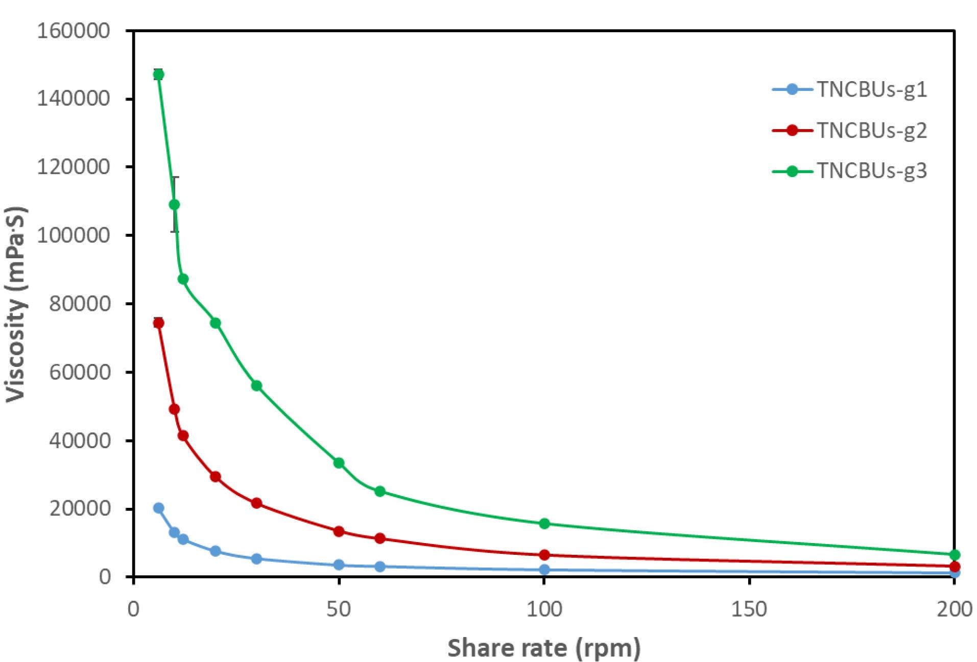
Figure 1.
Viscosity of TZN nanocubosomal gel formulations. (TNCUBs-g1 with 1% Carbopol in blue, TNCUBs-g2 with 1.5% Carbopol in red and TNCUBs-g3 with 2% Carbopol in green)
.
Viscosity of TZN nanocubosomal gel formulations. (TNCUBs-g1 with 1% Carbopol in blue, TNCUBs-g2 with 1.5% Carbopol in red and TNCUBs-g3 with 2% Carbopol in green)
Texture Analysis Results
Texture profile analysis revealed significant differences (P < 0.05) among the formulations. TNCUBs-g3 (2%) exhibited the highest firmness, TNCUBs-g2 (1.5%) demonstrated the greatest consistency with lowest adhesiveness, while TNCUBs-g1 (1%) displayed the highest cohesiveness (Figure S1 in supplementary data).
Drug Content
The drug content of the prepared TNCUBs gels (TNCUBs-g1, TNCUBs-g2, and TNCUBs-g3) ranged from 96.1% to 97.8%, with no statistically significant differences observed among the formulations (P > 0.05). These findings confirm the uniform incorporation and distribution of TZN within all gel preparations.
Selection of the Optimized TNCUBs-Gel Formulation
Based on texture profile, rheological characteristic, pH, and drug content, TNCUBs-g2 (1.5% Carbopol 934) was identified as the optimal formulation and selected for subsequent ex vivo, in vitro, stability, and in vivo studies.
In Vitro Drug Release
Figure 2 presents the in-vitro release profiles of the optimized TNCUBs-g2 and the TZN solution. The drug solution exhibited a rapid release pattern, achieving complete release within 2 h. In contrast, TNCUBs-g2 displayed a sustained release profile, with 59.7 ± 1.9% of TZN released over 24 h.
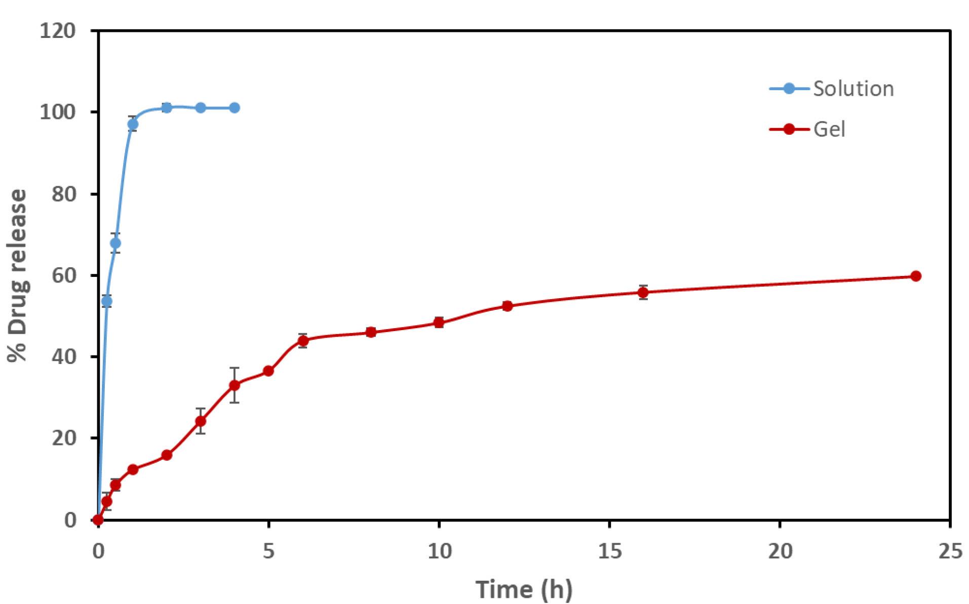
Figure 2.
In-vitro release profile of TZN solution and TNCUBs-g2 formulation in phosphate buffer (pH 7.4, 37 ± 1 °C). Data are presented as mean ± SD (n = 3)
.
In-vitro release profile of TZN solution and TNCUBs-g2 formulation in phosphate buffer (pH 7.4, 37 ± 1 °C). Data are presented as mean ± SD (n = 3)
Ex Vivo Permeation Study
The optimized TNCUBs-g2 gel exhibited a fourfold higher steady-state flux (32.32 µg/cm2/h) compared to the plain TZN solution (8.06 µg/cm2/h), along with a shorter lag time (0.83 ± 0.16 h vs. 2.3 ± 0.2 h) (Figure 3). Differences in flux values among all tested formulations were statistically significant (P < 0.05) (Table 2).
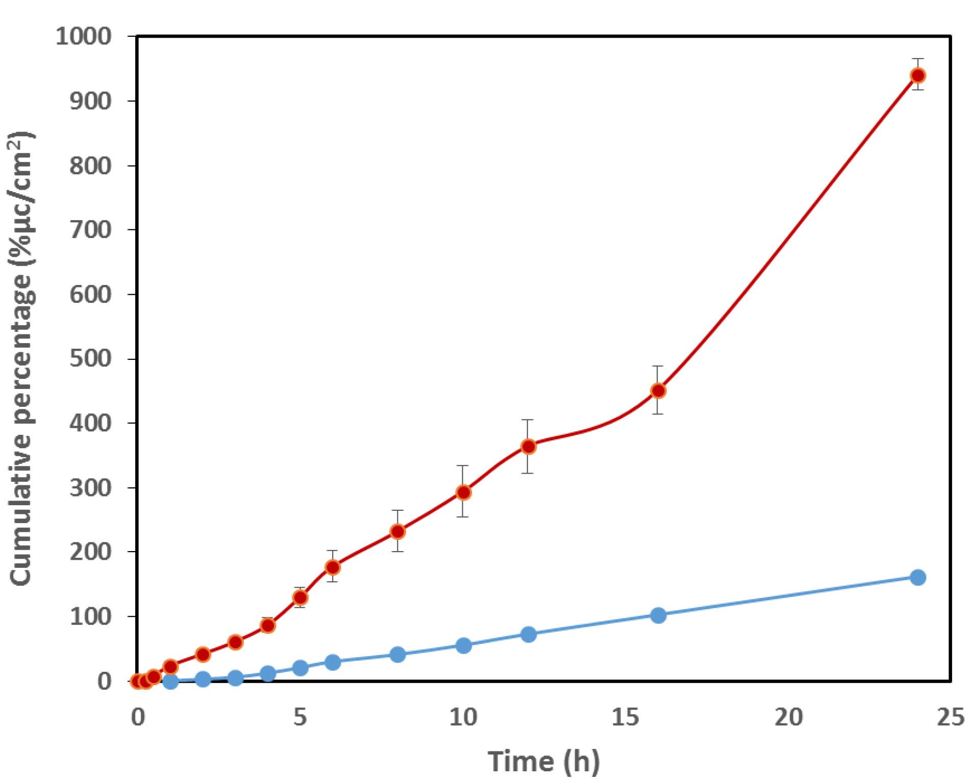
Figure 3.
Transdermal permeation profiles of TZN solution and the optimized TNCUBs-gel across rat skin over 24 h
.
Transdermal permeation profiles of TZN solution and the optimized TNCUBs-gel across rat skin over 24 h
Table 2.
Ex vivo permeation parameters of TZN solution and optimized TNCUBs-g2 gel across rat skin (Jss: steady-state flux; Papp: permeability coefficient).
|
Ex-vivo permeation parameters
|
Formulation
|
|
TZN solution
|
TNCUBs-gel
|
| Jss (μg/cm2 / h) |
8.061 ± 0.27 |
32.3 ± 1.6 |
| Papp (cm/h × 10-3) |
0.054 ± 0.06 |
0.220 ± 0.011 |
| Lag time (h) |
2.37 ± 0.2 |
0.829 ± 0.162 |
In Vivo Pharmacokinetics Study
The comparative bioavailability of the TNCUBs-gel and the oral TZN solution was evaluated in rats. The mean plasma concentration–time profiles are presented in Figure 4, and the corresponding pharmacokinetic parameters, calculated using PKSolver, are summarized in Table 3.
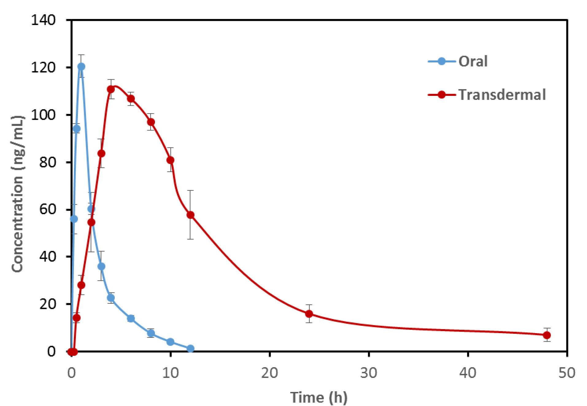
Figure 4.
Mean plasma concentration-time profile of oral TZN solution and transdermal TNCUBs-gel following administration of a 2.5 mg TZN dose
.
Mean plasma concentration-time profile of oral TZN solution and transdermal TNCUBs-gel following administration of a 2.5 mg TZN dose
Table 3.
pharmacokinetic parameters of TZN following transdermal administration of the nanocubosomal gel versus oral solution.
|
Pharmacokinetic Parameters
|
Oral-free TZN solution
|
TNCUBs-gel
|
| Cmax (ng/ml) |
120.53 ± 9.3 |
110.8 ± 4.1 |
| Tmax (h) |
2.0 ± 0.05 |
4.08 ± 0.07 |
| AUC0-24 ng.h/ml |
455.05 ± 9.5 |
1403.19 ± 42.1 |
| AUC0-∞ ng.h/ml |
458.24 ± 18.4 |
1549.2 ± 103.4 |
Pharmacokinetic parameters were determined using the PK-Solver software. Table 3 summarizes the results from triplicate measurements for both oral and transdermal administration routes. Statistical comparisons were conducted to identify significant differences between the two delivery methods.32
Statistical evaluation using the t-test revealed significant differences (P < 0.05) in pharmacokinetic parameters between the two administration routes. For the oral formulation, Cmax and Tmax were 120.53 ± 9.3 ng/mL and 2.0 ± 0.05 h, respectively. In contrast, the transdermal formulation exhibited Cmax = 110.8 ± 4.1 ng/mL and Tmax = 4.08 ± 0.07 h. The shorter Tmax and higher Cmax observed with the oral dose indicate rapid gastrointestinal absorption, whereas the transdermal system produced a delayed Tmax and slightly lower Cmax due to the diffusional resistance of the skin barrier effect, resulting in gradual systemic absorption.33
Moreover, the AUC₀–₂₄ for the transdermal application (1403.19 ± 42.1 ng·h/mL) was significantly higher than that of the oral solution (455.05 ± 9.5 ng·h/mL, P < 0.05), confirming sustained and enhanced drug exposure via transdermal delivery.
Discussion
The prepared TNCUBs-gel formulations displayed a uniform yellow color, smooth texture, and no evidence of phase separation, precipitation, or grittiness. These characteristics indicate good homogeneity, compatibility between the cubosomal dispersion and Carbopol polymeric matrix, and physical stability of the system.
The pH of all NCUBs gel formulations (7.4–7.6) was within the physiologically acceptable range for dermal applications, ensuring non-irritancy. Statistical analysis revealed no significant differences among gels with varying Carbopol concentrations (P > 0.05), indicating consistent formulation characteristic.
Rheological evaluation demonstrated that all TNCUBs gels (TNCUBs-g1, TNCUBs-g2, and TNCUBs-g3) exhibited non-Newtonian, shear-thinning (pseudoplastic) behavior, where viscosity decreased with increasing shear rate (6–200 rpm). Viscosity in Newtonian fluids remains constant regardless of the applied rotational force; however, the TNCUBs formulations demonstrated a consistent decline in viscosity as spindle speed increased.
Such flow behavior facilitates effortless application on the skin surface while preserving structural stability at rest. Increasing Carbopol 934 concentration from 1% to 2% significantly elevated viscosity, reflecting the formation of a denser polymeric network
The drug content of all TNCUBs gels ranged from 96.1% to 97.8%, complying with USP specifications (90–110%), and confirming efficient drug entrapment and uniform distribution. No significant differences were detected among gels with varying Carbopol concentrations (P > 0.05).
Texture profile analysis indicated that increasing Carbopol 934 concentration significantly influenced the mechanical properties of TNCUBs gels (P < 0.05). TNCUBs-g3 (2%) exhibited the highest firmness, while TNCUBs-g1 (1%) showed the greatest cohesiveness. Interestingly, TNCUBs-g2 (1.5%) presented the most balanced mechanical properties, highest consistency and lowest adhesiveness, suggesting optimal patient comfort during topical use.
As is presented in Figure 3, the in vitro release study demonstrated that TNCUBs-g2 exhibited markedly sustained release (~ 59.8% in 24 h) compared to the pure TZN solution (~101% in 2 h), confirming the prolonged drug release capacity of the cubosomal gel system. The improvement in flux can be attributed to the nanostructured architecture of cubosomes, which offer a large surface area, high drug-loading capacity, and intimate contact with the stratum corneum, thereby facilitating diffusion.34 Additionally, glyceryl monooleate (GMO), the lipid component of the vesicles, act as a penetration enhancer by fluidizing and disrupting the ordered lipid bilayers of the skin, leading to increased permeability.35 The significantly shorter lag time observed for the TNCUBs-gel (0.83 ± 0.16 h) compared to the plain drug solution (2.3 ± 0.2 h) further supports its enhanced permeation capability, likely due to hydration and loosening of the stratum corneum structure by cubosomal vesicles.36
The in vivo pharmacokinetic study confirmed that the transdermal TNCUBs-gel achieved a 3.1-fold increase in TZN bioavailability compared with the oral solution (P < 0.05), as evidenced by a higher AUC₀–∞, lower Cmax, and delayed Tmax, thereby confirming sustained percutaneous absorption. The enhanced bioavailability can be attributed to the combined effects of cubosomal structural and compositional features. The internal cubic nanostructure facilitates close interaction with the stratum corneum lipids and promotes drug permeation.37 GMO further contributes by disrupting the ordered skin lipid bilayers and enhancing membrane fluidity.38 Poloxamer, with its amphiphilic character, supports penetration by interacting with both aqueous and lipid domains of the skin.39 In addition, the nanoscale size and large surface area of cubosomes enable intimate skin contact, while their negative surface charge may transiently disturb the stratum corneum barrier, further promoting TZN diffusion.40,41 Importantly, the transdermal route circumvents physiological barriers such as first-pass hepatic metabolism, which commonly reduce the effective systemic dose following oral administration, thus contributing to the markedly enhanced bioavailability observed. Moreover, transdermal delivery provides controlled and sustained release, leading to more stable plasma concentrations, reduced dosing frequency, fewer systemic side effects, and improved patient compliance.42,43
Conclusion
The developed TNCUBs-g2 exhibited markedly higher relative bioavailability compared with the oral TZN solution. Therefore, this nanocubosomal gel represents a more convenient and effective alternative for transdermal administration of TZN. Future studies may further establish this transdermal delivery system as a promising approach to enhance TZN bioavailability and achieve sustained therapeutic effects.
Competing Interests
No conflict of interests was declared by the authors.
Ethical Approval
All animal experiments were conducted in accordance with institutional guidelines and approved by the Research Ethics Committee, College of Pharmacy, University of Baghdad, Iraq (Protocol No. REC02202506A).
Supplementary Files
Supplementary file 1 contains Figure S1.
(pdf)
Acknowledgements
The authors thank the Department of Pharmaceutics, College of Pharmacy, University of Baghdad for supporting the project.
References
- Nashat B, Al-Kinani K. Nanoemulsion Formulation of Lefl unomide for Transdermal Delivery: Preparation and Characterization. International journal of drug delivery technology 2023; 13:57-65. doi: 10.25258/ijddt.13.1.09 [Crossref] [ Google Scholar]
- Al-shaibani Ajn, Mowafaq MG. Polyvinyl Polymer- Based Olanzapine Nanoparticles for Transdermal Delivery: Design, In-vitro and Ex-vivo Evaluation. Iraqi Journal of Pharmaceutical Sciences 2025; 33(4SI):119-32. doi: 10.31351/vol33iss(4SI)pp119-132 [Crossref] [ Google Scholar]
-
Hasan MJ, Rajab NA. Nano vesicular topical drug delivery system: Types, structural components, preparation techniques, and characterizations. Journal of Emergency Medicine, Trauma and Acute Care 2024;2024(6 - Al-Bayan University Scientific Conference). doi: 10.5339/jemtac.2024.absc.16.
- Fadhel A, Ayash N. Tizanidine Nano emulsion: Formulation and in-vitro Characterization. Journal of Pharmaceutical Negative Results 2022; 13:572-81. doi: 10.47750/pnr.2022.13.03.086 [Crossref] [ Google Scholar]
- Gadad A, Patil A, Sariki VK, Dandagi P, Masthiholimath V. Design and characterization of biodegradable Tizanidine nanoparticles for improved oral bioavailability. Indian drugs 2016; 53:57-9. doi: 10.53879/id.53.03.10438 [Crossref] [ Google Scholar]
- Saba Abdulhadi J, Maha Mahdi A, Manar Adnan T, Omar Saeb S. Formulation and in-vitro evaluation of Tizanidine nanoparticle as a sublingual tablet. Journal of Advanced Pharmacy Education and Research 2025; 15(4):97-109. doi: 10.51847/N3pE1ycP4H [Crossref] [ Google Scholar]
- Sheikh F, Naaem Aamir M, Haseeb MT, Bukhari APDSNA, Farid-ul-Haq M, Akhtar N. Design, physico-chemical assessment and pharmacokinetics of a non-toxic orodispersible film for potential application in musculo-skeletal disorder. Journal of Drug Delivery Science and Technology 2021; 65:102726. doi: 10.1016/j.jddst.2021.102726 [Crossref] [ Google Scholar]
- Saini M, Kumar S, Jhawat V, Dass R. Design Strategies for Improving Oral Bioavailability of Tizanidine: Solid Dispersion Approach. INTERNATIONAL JOURNAL OF DRUG DELIVERY TECHNOLOGY 2024; 14:1708-15. doi: 10.25258/ijddt.14.3.65 [Crossref] [ Google Scholar]
- Jagdale S, Brahmane S, Chabukswar A. Optimization of Microemulgel for Tizanidine Hydrochloride. Antiinflamm Antiallergy Agents Med Chem 2020; 19(2):158-79. doi: 10.2174/1871523018666190308123100 [Crossref] [ Google Scholar]
- Farouk F, Khalil RM, Abdelbary A, El Arini SK, Basha M, El-Hashemy HA. Bioanalytical Comparison of Transdermal Delivery of Tizanidine from Different Nanovesicular Carriers. Journal of Pharmaceutical Innovation 2021; 16(2):384-90. doi: 10.1007/s12247-020-09447-z [Crossref] [ Google Scholar]
- Gade R, Rao CB, Ayanampudi A, Vegendla MR, Bhai VA, Nama S. Formulation and evaluation of mephenesin topical gel. World J Pharm Pharm Sci 2013; 2:1475-89. [ Google Scholar]
- Raheema DA, Kassab HJ. Preparation and in-vitro Evaluation of Secnidazole as Periodontal In-situ Gel for Treatment of Periodontal Disease. Iraqi Journal of Pharmaceutical Sciences 2022; 31(2):50-61. doi: 10.31351/vol31iss2pp50-61 [Crossref] [ Google Scholar]
- Thamer AK, Abood AN. Preparation and In vitro Characterization of Aceclofenac Nanosuspension (ACNS) for Enhancement of Percutaneous Absorption using Hydrogel Dosage Form. Iraqi Journal of Pharmaceutical Sciences 2021; 30(2):86-98. doi: 10.31351/vol30iss2pp86-98 [Crossref] [ Google Scholar]
- Cevher E, Sensoy D, Taha MAM, Araman A. Effect of Thiolated Polymers to Textural and Mucoadhesive Properties of Vaginal Gel Formulations Prepared with Polycarbophil and Chitosan. AAPS PharmSciTech 2008; 9(3):953-65. doi: 10.1208/s12249-008-9132-y [Crossref] [ Google Scholar]
- Patlolla VGR, Holbrook WP, Gizurarson S, Kristmundsdottir P. Evaluation of in vitro mucoadhesiveness and texture profile analysis of doxycycline in situ hydrogels. Die Pharmazie - An International Journal of Pharmaceutical Sciences 2020; 75(1):7-12. doi: 10.1691/ph.2020.9122 [Crossref] [ Google Scholar]
- Anjum F, Zakir F, Verma D, Aqil M, Singh M, Jain P. Exploration of Nanoethosomal Transgel of Naproxen Sodium for the Treatment of Arthritis. Current Drug Delivery 2020; 17(10):885-97. doi: 10.2174/1567201817666200724170203 [Crossref] [ Google Scholar]
- Omar Saeb S, Entidhar Jasim A-A. Pharmacokinetic parameters of ondansetron in rats after oral solution and transdermal invasomes gel: A comparison study. Journal of Advanced Pharmacy Education and Research 2023; 13(1):116-21. doi: 10.51847/HS5a27EI6o [Crossref] [ Google Scholar]
- Kulkarni GS, Sateesh Babu PR. Design and evaluation of tizanidine buccal mucoadhesive patches. J Appl Pharm Sci 2012; 2(4):74-81. doi: 10.7324/JAPS.2012.2411 [Crossref] [ Google Scholar]
- Ali T, Shoaib M, Yousuf R, Jabeen S, Muhammad I, Tariq A. Use of hydrophilic and hydrophobic polymers for the development of controlled release tizanidine matrix tablets. Brazilian Journal of Pharmaceutical Sciences 2014; 50:799-818. doi: 10.1590/S1984-82502014000400016 [Crossref] [ Google Scholar]
- Jacob S, Nair A, Morsy M. Dose Conversion Between Animals and Humans: A Practical Solution. Indian Journal of Pharmaceutical Education and Research 2022; 56:600-7. doi: 10.5530/ijper.56.3.108 [Crossref] [ Google Scholar]
- Al Abood RM, Talegaonkar S, Tariq M, Ahmad FJ. Microemulsion as a tool for the transdermal delivery of ondansetron for the treatment of chemotherapy induced nausea and vomiting. Colloids Surf B Biointerfaces 2013; 101:143-51. doi: 10.1016/j.colsurfb.2012.06.015 [Crossref] [ Google Scholar]
- Shin J-W, Seol I-C, Son C-G. Interpretation of animal dose and human equivalent dose for drug development. The Journal of Korean Oriental Medicine 2010; 31(3):1-7. [ Google Scholar]
- The National Committee for Research Ethics in Science and Technology. Guidelines for research ethics in science and technology; Norway 2009. The National Committee for Research Ethics in Science and Technology. Accessed September 5, 2025. Available from: https://www.forskningsetikk.no/en/guidelines/science-and-technology/.
- Taher SS, Kadhem Al-Kinani K. In vivo Brain Pharmacokinetics of Dolutegravir Sodium-Loaded Nanostructured Lipid Carrier in situ Gel: Comparative Study with an Intravenous Drug Solution. Al-Rafidain Journal of Medical Sciences 2025; 8(1):115-25. doi: 10.54133/ajms.v8i1.1692 [Crossref] [ Google Scholar]
- Sulaiman H, Ayash N. Soluplus and solutol hs-15 olmesartan medoxomil nanomicelle based oral fast dissolving film: In vitro and in vivo characterization. Farmacia 2024; 72:794-804. doi: 10.31925/farmacia.2024.4.7 [Crossref] [ Google Scholar]
- Kaviani R. Comparison of Different Approaches for Calculating LOD and LOQ in an HPLC-Based Analysis Method. Pharmaceutical Sciences 2024; 31(1):106-109. doi: 10.34172/PS.024.40452 [Crossref] [ Google Scholar]
- Dsouza L, Chaudhari P, Brahmam B, Lewis SA. Derma roller mediated transdermal delivery of tizanidine invasomes for the management of skeletal muscle spasms. European Journal of Pharmaceutical Sciences 2021; 165:105920. doi: 10.1016/j.ejps.2021.105920 [Crossref] [ Google Scholar]
- Alwan RM, A A. Rajab N Nanosuspensions of Selexipag: Formulation, Characterization, and in vitro Evaluation. Iraqi Journal of Pharmaceutical Sciences 2021; 30(1):144-53. doi: 10.31351/vol30iss1pp144-153 [Crossref] [ Google Scholar]
-
Abdulqader Aae, Sultan NAR. Preparation and characterization of Posaconazole as a Nano-micelles using d-α-Tocopheryl polyethylene glycol 1000 succinate (TPGS). Iraqi Journal of Pharmaceutical Sciences 2023;32(Suppl.):26–32. doi: 10.31351/vol32issSuppl.pp26-32.
- Hashim AA-J, A A. Rajab N A nastrozole Loaded Nanostructured Lipid Carriers: Preparation and Evaluation. Iraqi Journal of Pharmaceutical Sciences 2021; 30(2):185-95. doi: 10.31351/vol30iss2pp185-195 [Crossref] [ Google Scholar]
-
alik B, Eman BHA-K. Formulation and in vitro /in vivo Evaluation of Silymarin Solid Dispersion- Based Topical Gel for Wound Healing. Iraqi Journal of Pharmaceutical Sciences 2023;32(Suppl.):42–53. doi: 10.31351/vol32issSuppl.pp42-53.
- Kurji H, Ghareeb M, Zalzala M, Numan A, Hussain S. Serum Level Profile and Pharmacokinetic Parameters of Single Oral Dose of Metronidazole in Type II Diabetic Patients. Iraqi Journal of Pharmaceutical Sciences 2011; 20:18-23. doi: 10.31351/vol20iss1pp14-18 [Crossref] [ Google Scholar]
- Elshafeey AH, Kamel AO, Fathallah MM. Utility of nanosized microemulsion for transdermal delivery of tolterodine tartrate: ex-vivo permeation and in-vivo pharmacokinetic studies. Pharm Res 2009; 26(11):2446-53. doi: 10.1007/s11095-009-9956-5 [Crossref] [ Google Scholar]
- Pandian C, Sathali AA, Abirami G, Krithika E. Formulation development and characterization of triamcinolone loaded cubosomes for transdermal drug delivery. International Journal of Pharmaceutical Sciences and Medicine 2021; 6(12):1-17. doi: 10.47760/ijpsm.2021.v06i12.001 [Crossref] [ Google Scholar]
- Abdelbari MA, Elshafeey AH, Abdelbary AA, Mosallam S. Implementing Nanovesicles for Boosting the Skin Permeation of Non-steroidal Anti-inflammatory Drugs. AAPS PharmSciTech 2023; 24(7):195. doi: 10.1208/s12249-023-02649-x [Crossref] [ Google Scholar]
- Nithya R, Jerold P, Siram K. Cubosomes of dapsone enhanced permeation across the skin. Journal of Drug Delivery Science and Technology 2018; 48:75-81. doi: 10.1016/j.jddst.2018.09.002 [Crossref] [ Google Scholar]
- Esposito E, Cortesi R, Drechsler M, Paccamiccio L, Mariani P, Contado C. Cubosome dispersions as delivery systems for percutaneous administration of indomethacin. Pharm Res 2005; 22(12):2163-73. doi: 10.1007/s11095-005-8176-x [Crossref] [ Google Scholar]
- Kwon TK, Hong SK, Kim J-C. In vitro skin permeation of cubosomes containing triclosan. Journal of Industrial and Engineering Chemistry 2012; 18(1):563-7. doi: 10.1016/j.jiec.2011.11.031 [Crossref] [ Google Scholar]
- Yapar EA, Ýnal Ö. Poly(ethylene oxide)–Poly(propylene oxide)-Based Copolymers for Transdermal Drug Delivery: An Overview. Tropical Journal of Pharmaceutical Research 2013; 11(5):855-66. doi: 10.4314/tjpr.v11i5.20 [Crossref] [ Google Scholar]
- Al-Zheery WH, Kassab HJ. Pharmacokinetic Study of Oral Disulfiram Suspension and Topical Transdermal Nano-Invasomes Gel in Wistar Rats. Al-Rafidain Journal of Medical Sciences (ISSN 2789-3219) 2024; 7(1):159-63. doi: 10.54133/ajms.v7i1.1130 [Crossref] [ Google Scholar]
- Salah S, Mahmoud AA, Kamel AO. Etodolac transdermal cubosomes for the treatment of rheumatoid arthritis: ex vivo permeation and in vivo pharmacokinetic studies. Drug Deliv 2017; 24(1):846-56. doi: 10.1080/10717544.2017.1326539 [Crossref] [ Google Scholar]
- Tawfik MA, Eltaweel MM, Fatouh AM, Shamsel-Din HA, Ibrahim AB. Brain targeting of zolmitriptan via transdermal terpesomes: statistical optimization and in vivo biodistribution study by (99m)Tc radiolabeling technique. Drug Deliv Transl Res 2023; 13(12):3059-76. doi: 10.1007/s13346-023-01373-0 [Crossref] [ Google Scholar]
- Ali Ashoor J, M. Mohsin J, Mohsin HM, W. Mahde B, M. Gareeb M. Permeability Enhancement of Methotrexate Transdermal Gel using Eucalyptus oil, Peppermint Oil and Olive Oil. Iraqi Journal of Pharmaceutical Sciences 2022;30(Suppl.):16–21. doi: 10.31351/vol30issSuppl.pp16-21.