Pharmaceutical Sciences. 32(2):291-301.
doi: 10.34172/PS.026.43173
Original Article
A Molecular and Computational Study on the Effects of 5-Fluorouracil, and Dicyclomine on AGS Cells: Regulation of P53 and SNAIL2 Pathways
Fariba Ghodrati Conceptualization, Investigation, Methodology, Writing – original draft, 1 
Mahdi Bagheri Conceptualization, Data curation, Formal analysis, Methodology, Visualization, Writing – original draft, 2
Seyedeh Yasaman Kolki Data curation, Formal analysis, Validation, Writing – original draft, 3
Parham Motamedi Data curation, Formal analysis, Methodology, Software, Visualization, Writing – review & editing, 4
Elham Sadeghi Investigation, Validation, Writing – review & editing, 5
Hossein Ebrahimi Methodology, Resources, Supervision, Validation, Writing – review & editing, 6
Dhifaf Saleem Kareem Maliki Investigation, Resources, Software, 4
Hadis Musavi Conceptualization, Investigation, Project administration, Supervision, Writing – review & editing, 4, 7, *
Author information:
1Department of Biology, Science and Research Branch, Islamic Azad University, Tehran, Iran
2Department of Biological Science and Technology, Faculty of Nano and Bio Science and Technology, Persian Gulf University, Bushehr, Iran
3Department of Pharmacy, Islamic Azad University of Ayatollah Amoli Branch, Amol, Iran
4Pharmaceutical Sciences Research Center, Institute of Herbal Medicines and Metabolic Disorders, Mazandaran University of Medical Sciences, Sari, Iran
5Department of Anatomical Sciences, School of Medicine, Ilam University of Medical Sciences, Ilam, Iran
6Pharmaceutical Sciences Research Center, Faculty of Pharmacy, Mazandaran University of Medical Science, Sari, Iran
7Department of Clinical Biochemistry and Genetics, Faculty of Medicine, Mazandaran University of Medical Sciences, Sari, Iran
Abstract
Background:
5-Fluorouracil (5-FU) is commonly used to inhibit gastric cancer (GC) cell growth, but its clinical effectiveness is frequently limited by chemoresistance. Dicyclomine (DIC), an anticholinergic agent, has been suggested to enhance the therapeutic effect of 5-FU in cancer treatment.
Objectives:
This study aimed to investigate the effects of 5-FU, DIC, and their combination on AGS cell viability and the expression of SNAIL2 and P53.
Methods:
AGS cells were cultured in Dulbecco’s Modified Eagle Medium/F12 supplemented with 10% fetal bovine serum (FBS). Cell viability was assessed using the 3-(4,5-dimethylthiazol-2-yl)-2,5-diphenyltetrazolium bromide (MTT) assay. The mRNA expression levels of P53 and SNAIL2 were analyzed by quantitative real-time PCR (qRT-PCR). Drug synergism was evaluated using the CompuSyn software. Network pharmacology was employed to investigate the potential relationship between DIC and the expression of P53 and SNAIL2 genes.
Results:
The IC₅₀ values for 5-FU and DIC were 4 µg/mL and 800 µg/mL, respectively. In AGS cells, all treatments significantly upregulated P53 expression (5-FU: P<0.01; DIC: P<0.001; Combination: P<0.0001). While 5-FU (P<0.01) or DIC (P<0.0001) alone increased SNAIL2 expression, their combination significantly reduced it (P<0.05). CompuSyn software confirmed a synergistic interaction. KEGG pathway analysis revealed 38 enriched pathways, with the adherens junction pathway, highlighted due to its potential role in SRC protein-mediated downregulation of SNAIL gene expression.
Conclusion:
These findings indicate that the combination of 5-FU and DIC synergistically exerts significant anticancer effects by upregulating P53 and downregulating SNAIL2 expression, representing a promising strategy for optimizing GC management.
Keywords: Gastric neoplasms, TP53, SNAIL2, Fluorouracil, Dicyclomine, Drug synergism
Copyright and License Information
© 2026 The Author(s).
This is an open access article and applies the Creative Commons Attribution Non-Commercial License (
http://creativecommons.org/licenses/by-nc/4.0/). Non-commercial uses of the work are permitted, provided the original work is properly cited.
Funding Statement
No funding.
Introduction
GC is one of the most prevalent cancers worldwide, arising from a complex interplay of environmental and genetic factors1. The risk of developing GC is predominantly driven bymodifiable lifestyle factors including dietary habits, physical inactivity, alcohol consumption, smoking, and H. pylori infection.2 Despite notable advances in therapeutic approaches over recent years, managing and effectively treating gastric cancer remains a significant clinical challenge due to the disease’s aggressive nature and the emergence of drug resistance.3 One of the primary chemotherapeutic agents used to inhibit gastric cancer growth is 5-FU. However, the clinical utility of 5-FU is often limited by chemo-resistance, which reduces its efficacy and leads to treatment failure.4 Combining 5-FU with adjuvant agents is an established strategy to overcome chemoresistance, with compounds such as tamoxifen,5 curcumin,6 and glabridin7 being explored in this context. While the concept of 5-FU combination therapy is well-established, the novelty of the present study lies in its investigation of DIC as a novel sensitizing agent that targets muscarinic receptor signaling. Although traditionally prescribed for irritable bowel syndrome, DIC has recently demonstrated potential anti-cancer properties due to its action as a muscarinic receptor antagonist.8 DIC has been shown to restore chemosensitivity in prostate cancer by blocking the muscarinic receptor M1(M1R). In both cell and animal models, combining DIC with docetaxel reverses chemotherapy resistance, enhancing the drug’s cytotoxic effects. This combination highlights the pivotal role of M1R in mediating chemoresistance, establishing DIC as a promising adjuvant to enhance the efficacy of docetaxel in resistant prostate cancer cases.9 The broader relevance of this mechanism is supported by growing evidence that muscarinic acetylcholine receptors (mAChRs) actively contribute to oncogenesis, influencing key cancer hallmarks such as proliferation, evasion of apoptosis, and migration through diverse signaling pathways.10
This pathway holds particular significance in the digestive tract, where autocrine and paracrine acetylcholine signaling through mAChRs fosters a tumor-promoting microenvironment. In gastric cancer, the M3 muscarinic receptor (M3R) is a key driver of cell proliferation, mechanistically linked to pro-tumorigenic pathways including β-catenin signaling. Acetylcholine acts primarily through M3R to activate downstream signaling cascades such as EGFR/ERK and AKT, which in turn promote gastric cancer cell growth. Selective M3R antagonists like 4-DAMP and darifenacin effectively suppress tumor growth in vitro and in vivo by inhibiting these pathways. Additionally, blocking M3R enhances the sensitivity of gastric cancer cells to chemotherapeutic agents such as 5-FU, inducing apoptosis and diminishing anti-apoptotic protein expression.11
At the molecular level, the tumor suppressor gene p53 is a central regulator in GC pathogenesis. Mutations in TP53 rank among the most frequent genetic alterations in GC, with reported prevalence varying widely from approximately 37% to 77%, influenced by tumor stage and population studied.12 These mutations disrupt p53’s crucial role in maintaining genomic stability, thereby facilitating tumor initiation and progression. Notably, hotspot mutations such as R175 and R248 correlate with increased aggressive phenotypes, and poorer overall survival in GC patients.13,14 Loss of p53 function promotes malignant transformation by enabling epithelial-mesenchymal transition (EMT), enhancing cancer stemness, and impairing programmed cell death mechanisms.15 Chronic inflammatory insults, including Helicobacter pylori infection, exacerbate this effect by inhibiting p53-dependent DNA repair pathways, thus accelerating carcinogenesis.16 Furthermore, the chemotherapeutic agent 5-FU exerts part of its anti-tumor activity by inducing p53-dependent cell cycle arrest and apoptosis.17 However, mutated p53 in cancer stem cells (CSCs) confers resistance to 5-FU, diminishing treatment effectiveness and contributing to tumor recurrence.18
Another critical molecule involved in GC aggressiveness and chemoresistance is SNAIL2, a member of the SNAIL family of transcription factors. SNAIL2 (also known as SNAI2) is intricately linked to poor clinical outcomes by promoting EMT, a key biological process enabling tumor invasion and metastasis.19 It functions by repressing the epithelial marker E-cadherin and upregulating mesenchymal markers, thereby facilitating cellular plasticity critical for metastatic dissemination.20 The role of SNAIL extends to maintaining the CSC phenotype in cancers, which is a major driver of therapeutic resistance.21-23 Through EMT-mediated mechanisms, SNAIL2 enhances CSC properties, including survival and self-renewal. CSCs exhibit high drug efflux capacity via overexpression of ATP-binding cassette (ABC) transporters, particularly contributing to resistance against chemotherapeutic agents.24,25 Moreover, SNAIL2 supports evasion of apoptosis, further highlighting its significance in tumor progression and treatment failure.This study aimed to investigate the effects of 5-FU, DIC, and their combination on the viability of AGS cells, as well as on the expression of the tumor suppressor P53 and the EMT-related transcription factor SNAIL2.
Methods and Materials
Experimental Study
Cell culture
The human gastric adenocarcinoma AGS cell line (Pasteur Institute, Tehran, Iran) was cultured in Dulbecco’s Modified Eagle Medium/Nutrient Mixture F-12 (DMEM/F-12) (Sigma-Aldrich, USA) supplemented with 10% heat-inactivated fetal bovine serum (FBS), 100 μg/ml glutamine, 100 U/mL penicillin, and 100 µg/mL streptomycin (Sigma-Aldrich, USA). Cells were maintained in a humidified incubator at 37°C with 5% CO₂.
MTT Assay
Cell viability was assessed using the standard MTT assay. Briefly, AGS cells were seeded in 96-well plates at 5 × 10³ cells/well in complete DMEM/F12 medium containing 10% FBS at 37°C under 5% CO₂. After reaching 80% confluence, cells were treated with 5-FU (dissolved in PBS as a vehicle: 0.1, 0.5, 1, 2, and 4 µg/mL) and DIC (dissolved in DMSO as vehicle: 100, 300, 400, 600, 800, and 1000 µg/mL) for 48 hours, or combination treatments using serial dilutions (20-80%) of the IC₅₀ concentrations of 5-FU and DIC.
To assess the synergistic effect of 5-FU and DIC, a fixed-ratio combination based on the individual IC₅₀ values of each drug was prepared and tested. The procedure was as follows: A primary stock solution was prepared such that it simultaneously contained the pre-determined IC₅₀ concentration of 5-FU and the pre-determined IC₅₀ concentration of DIC in the culture medium. This primary combination stock was considered the 100% concentration point. From this stock, a series of sequential dilutions was prepared in culture medium to yield final working solutions with relative concentrations of 90%, 80%, 70%, 60%, 50%, 40%, 30%, 20%, and 10% of the original primary stock. This dilution scheme ensured that the proportional ratio between 5-FU and DIC remained constant across all tested concentration points. Cells were then treated with these serial dilutions for 48 hours. Following the treatment period, cell viability was assessed using the MTT assay. In this assay, 100 µL of MTT solution (5.0 mg/mL) was added to each well, and the plates were incubated for 4 hours at 37°C. Subsequently, 175 μL of DMSO was added,and the plates were shaken for 15 minutes to ensure complete dissolution. Absorbance was measured at 570 nm using an ELISA reader (ELX800, BioTek, USA). A new dose-response curve for the drug combination was generated, and the combination IC₅₀ defined as the relative concentration of the combination that inhibited 50% of cell viability was calculated. Cell viability percentages were calculated relative to the untreated control group, and all experiments were performed in triplicate.
Real-Time PCR
Total RNA was extracted using Trizol reagent (GeneAll, Korea) according to the manufacturer’s instructions. The RNA concentration and purity were assessed by measuring the optical density ratio (A260/A280) with a NanoDrop 1000 spectrophotometer (Wilmington, DE, USA). Complementary DNA (cDNA) synthesis was performed using the Two-Step cDNA Synthesis Kit (GeneAll, Korea) according to the manufacturer’s protocol. Quantitative real-time PCR (qPCR) was carried out using SYBR Green-based Master Mix (Amplicon kit, Denmark) (ABI 7500 thermal cycler, Applied Biosystems, Foster City, CA, USA). Primer sequences used for gene expression analysis are listed in Table 1. All samples were run in triplicate. Gene expression levels were quantified using the Pfaffl method, which is an efficiency corrected model for relative quantification. Cycle threshold (Ct) values were normalized against GAPDH expression as the internal control. For the qPCR analysis, RNA was extracted from three independent biological replicates per treatment group (n = 3), with each biological sample analyzed in technical triplicate. The specificity of the amplification for each primer set was confirmed by melt-curve analysis after each qPCR run.
Table 1.
Primer sequences
|
Genes Name
|
Primers
|
Sequences
|
|
GAPDH
|
forward |
5'-ACTCTGGTAAAGTGGATATTGTTGC -3' |
| reverse |
5'-GGAAGATGGTGATGGGATTTC -3' |
|
SNAIL2
|
forward |
5'- ATTCGGACCCACACATTACC -3' |
| reverse |
5'- GCAGTGAGGGCAAGAAAAAG -3' |
|
P53
|
forward |
5'-TAACAGTTCCTGCATGGGCGGC-3' |
| reverse |
5'-AGGACAGGCACAAACACGCACC-3' |
Synergistic Effects Analysis
Quantitative assessment of drug synergism was performed using CompuSyn software (ComboSyn, Inc.). The combination index (CI) was calculated using the following equation:
CI = (D₁ / (Dₓ)₁) + (D₂ / (Dₓ)₂)
where D₁ and D₂ are the concentrations of 5-FU and DIC in the combination required to achieve an x% effect, while (Dₓ)₁ and (Dₓ)₂ are the concentrations of each drug alone required to produce the same effect.
Furthermore,the Fa-CI plot, dose-effect curve, and isobologram analyses were generated. The Fa-CI plot illustrates the actual CI values at various suppression levels. The Dose-Reduction Index (DRI) was also calculated to determine the extent of dose reduction for each drug in a synergistic combination. A DRI value greater than 1 indicates a favorable dose reduction for that drug in the combination, whereas a value less than 1 suggests an unfavorable reduction. A value of 1 denotes no dose reduction.
Network Pharmacology Analysis and Identification of Biological Targets for Dicyclomine, Gastric Cancer, and 5-FU Resistance
The Simplified Molecular Input Line Entry System (SMILES) notation of DIC was retrieved from PubChem and used as an input for the Swiss Target Prediction database to predict its potential human protein targets. Simultaneously, genes associated with gastric cancer (GC) were obtained from the Online Mendelian Inheritance in Man (OMIM) and GeneCards databases. Venn diagrams, generated using an online tool (http://bioinformatics.psb.ugent.be/webtools/Venn/), were then employed to identify the common targets between the predicted DIC targets, GC-related targets, and genes associated with 5-FU resistance.
The Protein-Protein Interaction Network
To identify protein-protein interactions among the common targets of DIC, GC-related targets, and 5-FU resistance targets, the STRING database (https://string-db.org/) was used. The analysis was performed using 22 gene entries from Homo sapiens, with a high confidence interaction score threshold set at > 0.7. The resulting protein-protein interaction (PPI) data were visualized and analyzed using Cytoscape version 3.9. The CytoHubba plugin within Cytoscape was then used to identify the key targets of DIC against GC, applying the Maximal Clique Centrality (MCC) algorithm.
Enrichment Analysis of Gene Ontology (GO) and Kyoto Encyclopedia of Genes and Genomes (KEGG)
To obtain information about gene ontology and to identify the canonical pathways, biological processes (BPs), cellular components (CCs), and molecular functions (MFs) associated with the target genes, we used the Annotation, Visualization, and Integrated Discovery (DAVID) database (https://david.ncifcrf.gov/). Subsequently, a graphical representation of the results was generated using tools available on using tools available at the Bioinformatics platform (http://www.bioinformatics.com.cn), which facilitated the creation of a bubble plot.
Dicyclomine-Target-Pathway Network
The dicyclomine-target-pathway network was constructed by combining the pathway networks obtained from the KEGG database and the dicyclomine-gastric cancer target network using Cytoscape software version 3.9.
Statistical Analysis
Data were analyzed and presented as mean ± SD. using GraphPad Prism statistical software (version 8.0; San Diego, CA, USA). Student’s t-test or one-way ANOVA followed by a post hoc test was used to compare the groups. Exact P values are reported in the Results section. Differences were considered statistically significant at P ≤ 0.05.
Results
Cytotoxicity Analysis
MTT assays revealed that cell viability was suppressed by 5-FU in a dose-dependent manner (P < 0.05). As shown in Figure 1A and 1B, the IC₅₀ values of 5-FU and DIC were 4 μg/mL and 800 μg/mL, respectively. The combination treatment synergistically reduced cell viability compared to either 5-FU or DIC monotherapy, achieving statistical significance (P < 0.05).
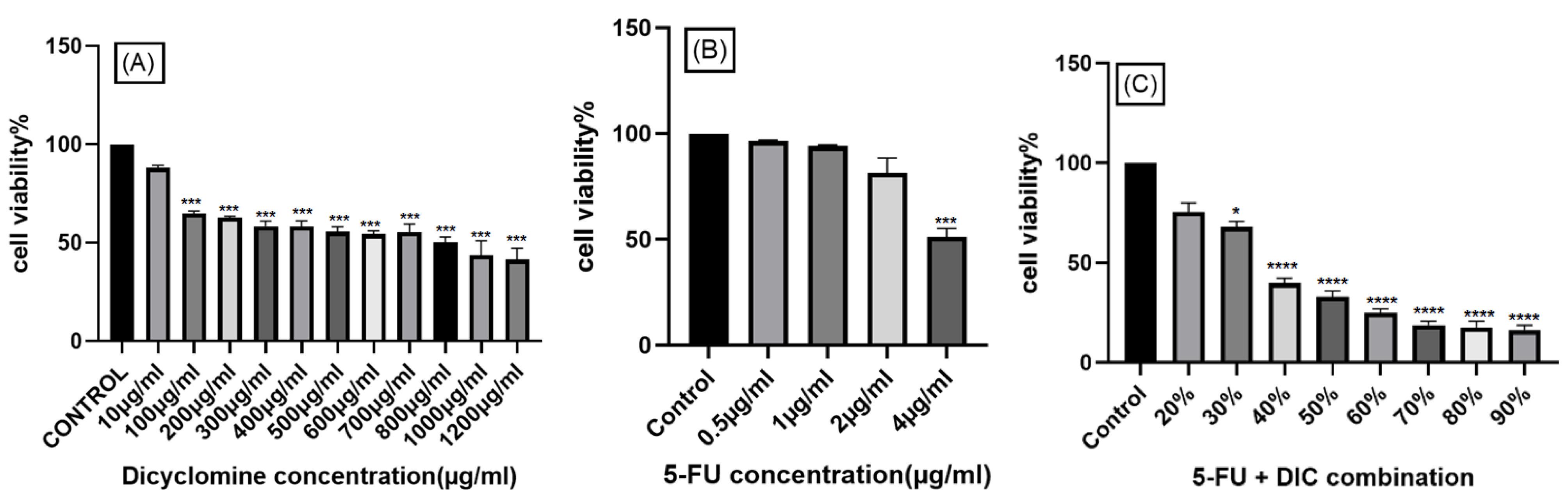
Figure 1.
Comparison of cell viability in AGS cell lines treated with (A) DIC, (B) 5-FU, and (C) the combination of 5-FU and DIC at the 48 h time point. The result are shown as the mean ± SD (n = 3). Significant differences compared to the control group are indicated as *P < 0.05, ***P < 0.001, and ****P < 0.0001
.
Comparison of cell viability in AGS cell lines treated with (A) DIC, (B) 5-FU, and (C) the combination of 5-FU and DIC at the 48 h time point. The result are shown as the mean ± SD (n = 3). Significant differences compared to the control group are indicated as *P < 0.05, ***P < 0.001, and ****P < 0.0001
Effect of 5-FU, DIC and 5-FU + DIC on P53 and SNAIL2 Gene Expression
To investigate the therapeutic mechanisms of 5-FU, DIC, and their combination, we analyzed the expression of the key regulatory genes P53 and SNAIL2 in AGS cells. As shown in Figure 2, P53 expression was significantly upregulated in the 5-FU (P < 0.01), DIC (P < 0.001), and 5-FU + DIC (P < 0.0001) treatment groups compared to the control. Furthermore, the upregulation in the combination group was significantly greater than that caused by either 5-FU or DIC alone (P < 0.0001). In contrast, SNAIL2 expression was significantly upregulated by 5-FU (P < 0.01) and DIC (P < 0.0001) alone, but was significantly downregulated in the 5-FU + DIC combination group compared to the single-agent treatments (P < 0.05).
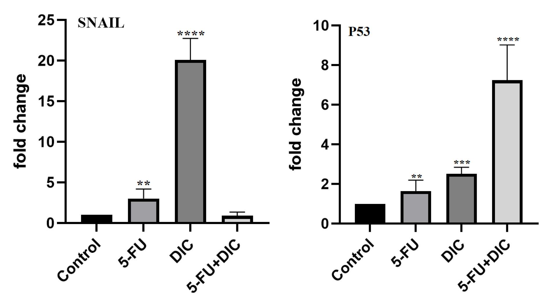
Figure 2.
Comparison of the effects of 5-FU, DIC, and 5-FU + DIC treatment on P53 and SNAIL2 gene expression. The results are shown as the mean ± SD (n = 3); Significant differences compared to the control group are indicated as **P < 0.01, ***P < 0.001, and ****P < 0.0001
.
Comparison of the effects of 5-FU, DIC, and 5-FU + DIC treatment on P53 and SNAIL2 gene expression. The results are shown as the mean ± SD (n = 3); Significant differences compared to the control group are indicated as **P < 0.01, ***P < 0.001, and ****P < 0.0001
Drug Interaction Analysis of 5-FU, DIC and 5-FU + DIC
Combination drug therapies can be either synergistic (more effective together), antagonistic (less effective), or additive (as effective as their individual effects combined).26 The efficacy of various DIC and 5-FU concentrations, used at a fixed ratio of 200:1 for 48 hours, was assessed to systematically evaluate the therapeutic potential of their co-administration. Different methods, including isobologram analysis, combination index (CI), and curve-shift analysis, were used to investigate the combined effect of both drugs. Dose-response analysis confirmed the beneficial effects of the combination treatment, which indicated that 5-FU and DIC are more effective when combined compared to either agent alone (Figure 3A). Examining the ‘m’ values (the slope of the median-effect plot) showed a sharper curve for the combination of 5-FU and DIC compared to either drug alone (Table 2 and Figure 3B). While dicyclomine’s significant linear correlation (R2 > 0.94) complies with the median-effect principle (r ≥ 0.95), 5-FU displays lower values of ‘r’ and ‘R2’. However, the combination of the two agents demonstrates a strong linear correlation, confirming the effectiveness of the combination (Table 2). CompuSyn analysis demonstrated that combining DIC and 5-FU synergistically suppressed the growth of AGS cells (Figure 3C-D). CI values ≤ 1 (Table 3) indicate synergy.27 Table 2 shows synergistic effects at Fa = 0.1, 0.25, and 0.5, as all CI values are ≤ 1. The findings demonstrate that the 5-FU and DIC combination inhibited the growth of AGS cells. The DRI values > 1 (Figure 4) indicate that combining 5-FU and DIC causes significant dose reductions while maintaining therapeutic efficacy.
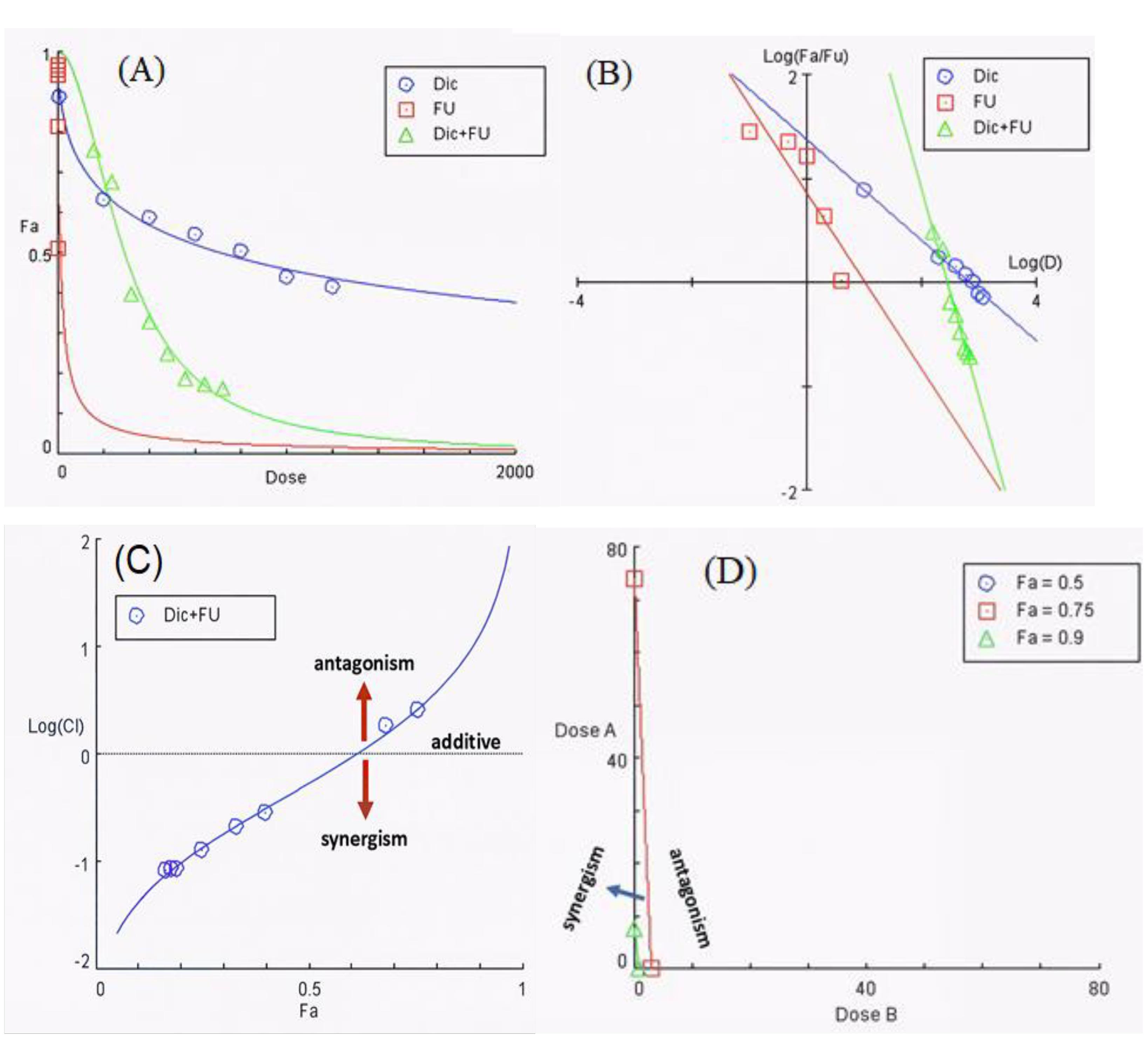
Figure 3.
The combination effects of DIC and 5-FU over 48 h. Panel A-B show the dose-response curve and the median-effect plot respectively, where the slope (m), the correlation coefficient (r) and R2 (r squared) describe the relationship between drug dose and effect. Panel C presents CI plots showing synergy (CI < 1), antagonism (CI > 1), and additive effects (CI = 1). Panel D highlights the isobologram depicting synergistic dose pairs
.
The combination effects of DIC and 5-FU over 48 h. Panel A-B show the dose-response curve and the median-effect plot respectively, where the slope (m), the correlation coefficient (r) and R2 (r squared) describe the relationship between drug dose and effect. Panel C presents CI plots showing synergy (CI < 1), antagonism (CI > 1), and additive effects (CI = 1). Panel D highlights the isobologram depicting synergistic dose pairs
Table 2.
The quantity of m (slope) and R2 (is the square of the correlation coefficient (r)) for medicines.
|
|
m
|
R2
|
| 5-FU |
-0.8 |
0.73 |
| DIC |
-0.4 |
0.98 |
| Combination of DIC and 5-FU (200:1) |
-2.01 |
.96 |
Table 3.
Affected fractions (Fa), CI (combination index) and DRI (the dose-reduction index) values calculated by CompuSyn software for determining the drug combination level. The CI > 1, CI = 1, and CI < 1 respectively show antagonism, additive, and synergism. DRI = 1 indicates dose doesn't reduce, whereas DRI > 1 and DRI < 1 show favorable and unfavorable dose reduction, respectively. CI values < 1 at Fa = 0.1–0.5 indicate synergism.
|
Fa (Effect)
|
Dose (5-FU)
(µg/mL)
|
Dose (DIC)
(µg/mL)
|
DRI
(5-FU)
|
DRI
(DIC)
|
CI Value
|
| .10 |
143.158 |
67139.4 |
33.0697 |
77.5466 |
0.04313 |
| .25 |
39.572 |
6932.35 |
15.5749 |
13.8163 |
0.13658 |
| .50 |
10.6657 |
715.847 |
7.33533 |
2.46163 |
0.54256 |
| .75 |
2.91121 |
73.9166 |
3.45473 |
0.43585 |
2.56953 |
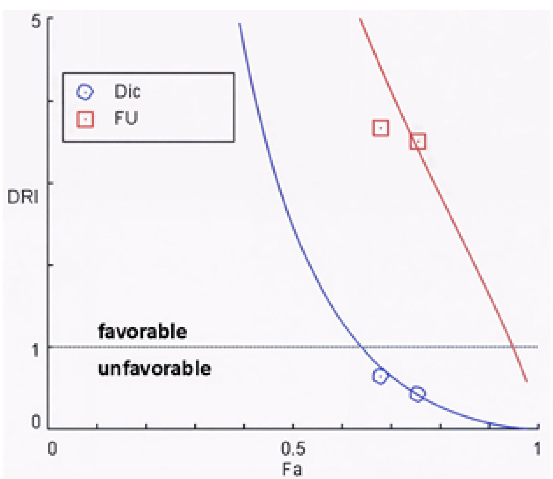
Figure 4.
Displays DRI values, with DRI > 1 indicating beneficial dose reductions. All results were generated using CompuSyn
.
Displays DRI values, with DRI > 1 indicating beneficial dose reductions. All results were generated using CompuSyn
Identification of Common Targets of DIC and Gastric Cancer
To identify common potential therapeutic targets of DIC in GC, we conducted a comprehensive bioinformatics analysis using the Swiss Target Prediction, GeneCards, and OMIM databases. The Swiss Target Prediction analysis yielded 106 targets for DIC, while the GeneCards and OMIM databases identified 2118 GC-related targets and 93 5-FU resistance-related targets, respectively. A Venn diagram analysis revealed four shared targets among all three groups and 18 shared targets between DIC and GC targets (Figure 5A).
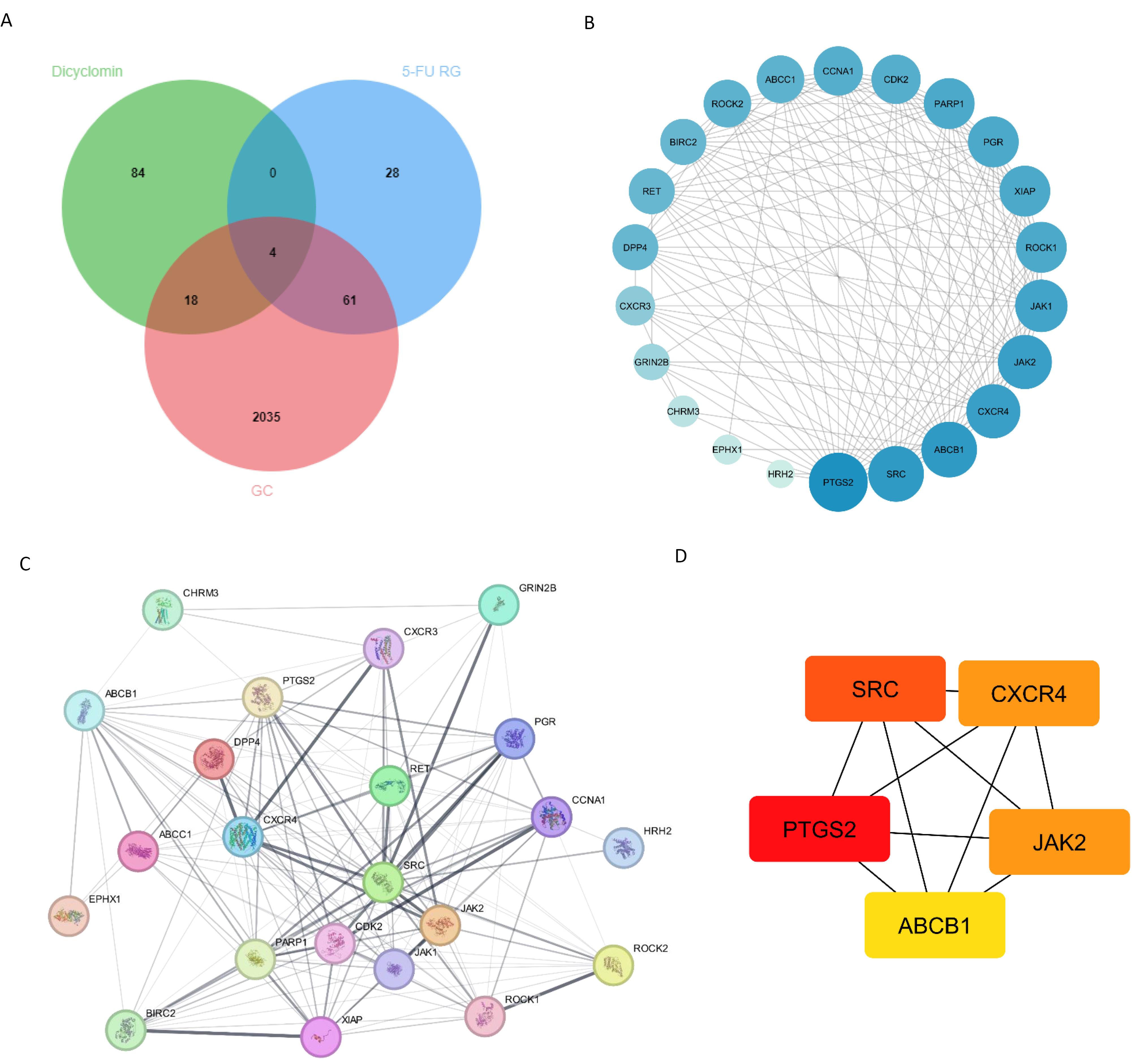
Figure 5.
A) Venn diagram illustrating the common targets between diclomine, gastric cancer (genes, and 5-FU resistant genes (5-FU RG) B) PPI network diagram of potential targets visualized in cytoscape software (larger size and bold color indicate more interactions) C) The PPI network was constructed using the STRING database. D) Five key targets in this network were identified using the CytoHubba plugin in Cytoscape. Darker colors indicate more critical targets
.
A) Venn diagram illustrating the common targets between diclomine, gastric cancer (genes, and 5-FU resistant genes (5-FU RG) B) PPI network diagram of potential targets visualized in cytoscape software (larger size and bold color indicate more interactions) C) The PPI network was constructed using the STRING database. D) Five key targets in this network were identified using the CytoHubba plugin in Cytoscape. Darker colors indicate more critical targets
Construction of Protein-Protein Interaction Network and Identification of Hub Genes
We constructed a PPI network of the 22 common targets using STRING and visualized it using Cytoscape (version 3.6.0). The network comprised 22 nodes and 149 edges, with an average number of neighbors of 13.545 and a local clustering coefficient of 0.852 (Figure 5B and Figure 5C). By employing the MCC algorithm in CytoHubba, we identified ten key hub genes, including JAK2, PTGS2, ABCB1, CXCR4, and SRC (Figure 5D). In the network visualization, node size and color intensity were scaled according to their degree of connectivity, with larger diameters and darker hues representing higher-degree nodes.
Functional Enrichment Analysis and Compound-Targets-Pathways Network
To elucidate the potential pharmacological mechanism of DIC in GC, we conducted GO and KEGG pathway analyses on the 22 common targets using the clusterProfiler package. The analysis identified 100 BP, 20 CCs, and 34 MF terms with P < 0.05 and count > 2. The top 10 GO results are displayed in Figure 6A. KEGG enrichment analysis revealed that dicyclomine’s mechanism against GC involves 11 targets, including XIAP, JAK2, CDK2, ROCK2, CXCR4, PTGS2, BIRC2, CCNA1, ROCK1, RET, and JAK1. Among these pathways, the adherens junction pathway emerged as a key pathway linking DIC to the SNAIL gene via SRC and ROCK proteins (Figure 6B and Figure 7). To further investigate the interactions between DIC, its targets, and associated pathways, we constructed a dicyclomine-target-pathway network diagram (Figure 6C). This network included 35 nodes, comprising the 22 common targets, 12 enriched KEGG pathways, and the active compound (DIC). The network contained 84 edges. SRC, XIAP, ROCK1, BIRC2, and ROCK2 were identified as the top targets based on their degree values.
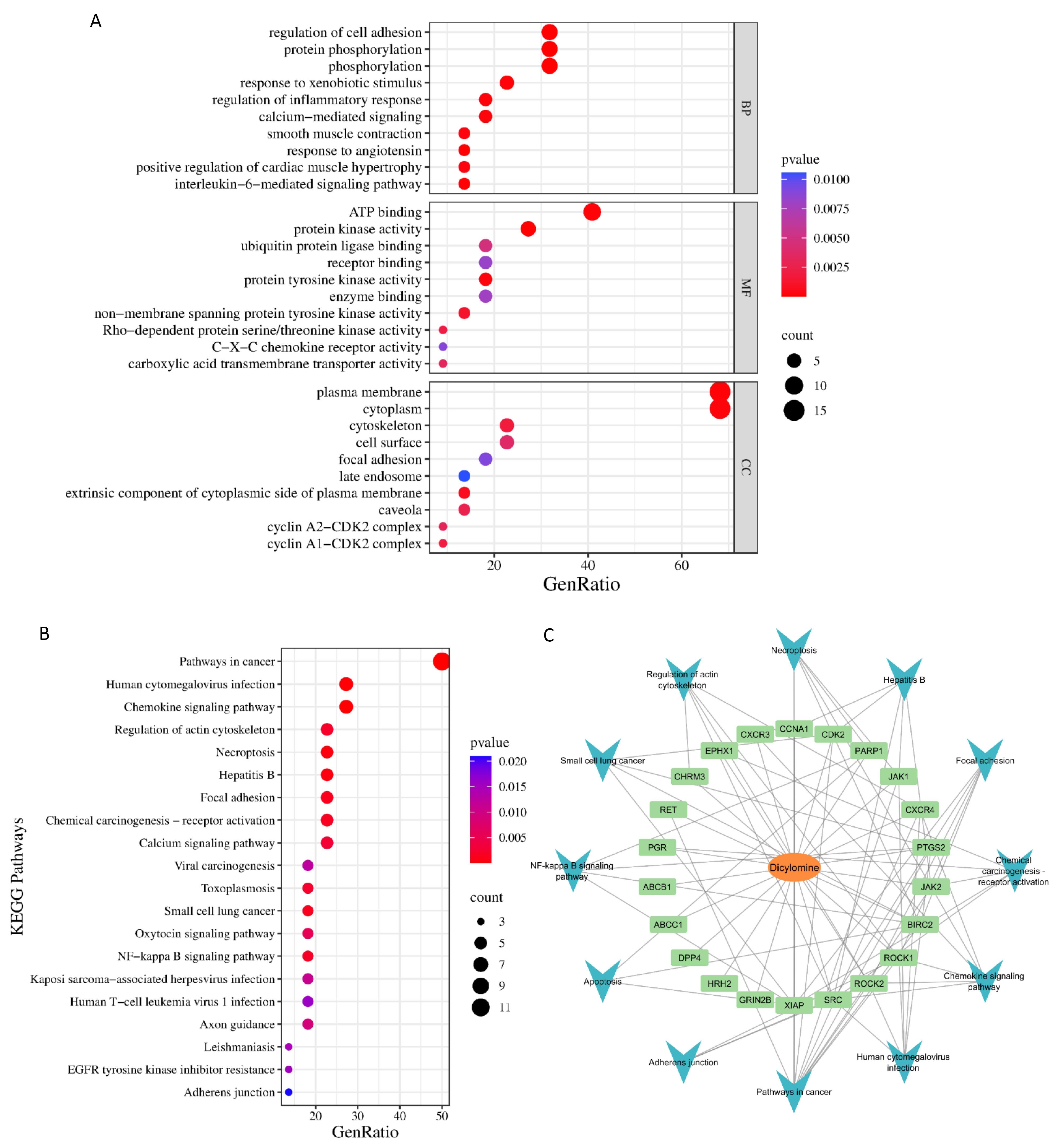
Figure 6.
A) Gene Ontology (GO) – Functional Enrichment Analysis, including the top 10 results from Biological Process (BP), Molecular Function (MF), and Cellular Component (CC). B) KEGG pathway analysis identified the top 20 enriched pathways among the 22 selected targets. C) “Compound-Pathway-Target” network diagram. The middle layer represents the targets and the outer layer represents the cancer related pathways
.
A) Gene Ontology (GO) – Functional Enrichment Analysis, including the top 10 results from Biological Process (BP), Molecular Function (MF), and Cellular Component (CC). B) KEGG pathway analysis identified the top 20 enriched pathways among the 22 selected targets. C) “Compound-Pathway-Target” network diagram. The middle layer represents the targets and the outer layer represents the cancer related pathways
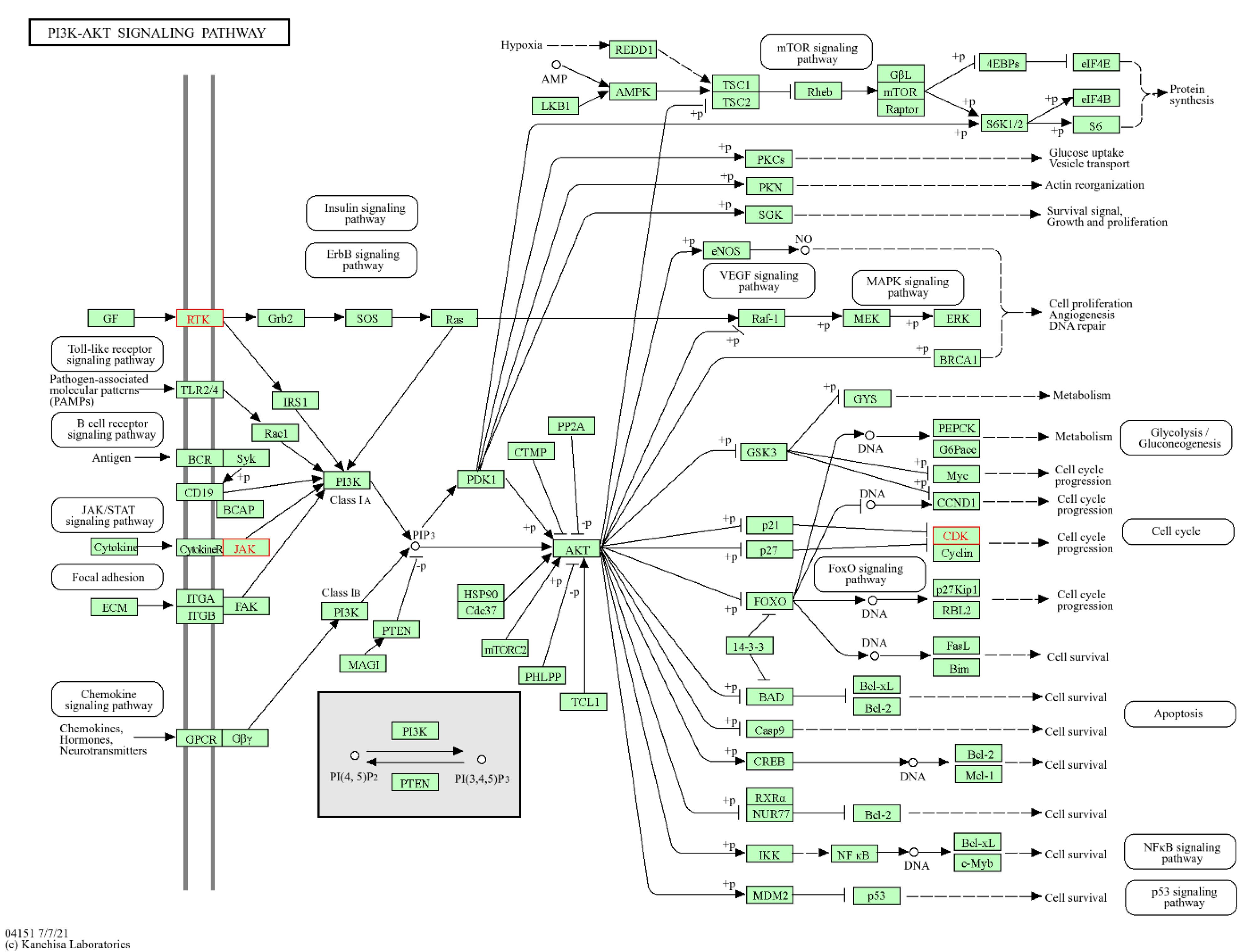
Figure 7.
Impact of dicyclomine on the adherens junction signaling pathway. Targets highlighted in red denote proteins modulated by dicyclomine within this pathway
.
Impact of dicyclomine on the adherens junction signaling pathway. Targets highlighted in red denote proteins modulated by dicyclomine within this pathway
Discussion
The present study suggests that combination therapy involving 5-FU and DIC may enhance the therapeutic response in GC. Treatment with 5-FU alone induced a clear dose-dependent reduction in AGS cell viability, with an IC₅₀ value of 4 µg/mL. This finding aligns with previous studies reporting the cytotoxic and antiproliferative effects of 5-FU in various cancer cell lines, including GC cells.28-30 Notably, co-treatment with 5-FU and DIC produced a more pronounced decrease in cell viability compared with either agent alone, indicating a potential synergistic interaction (P ≤ 0.05). CompuSyn analysis supported this observation through a lower IC₅₀ value for the combination. However, the relatively high IC₅₀ of DIC (800 µg/mL) suggests limited intrinsic cytotoxicity, which could limit its clinical utility despite its modulatory effect on 5-FU activity.DIC, an anticholinergic drug that selectively blocks the M1 muscarinic receptor, has been explored for potential anticancer properties. Previous research indicates that muscarinic receptor antagonism can suppress tumor cell proliferation and sensitize resistant cancer cells to chemotherapy.31 The M1 receptor, expressed in both normal and malignant cells, activates pro-survival pathways such as MAPK and PI3K/Akt when stimulated. Therefore, its inhibition could reduce pro-tumorigenic signaling and promote apoptosis. In gastrointestinal cancers, muscarinic signaling contributes to a tumor-supportive microenvironment by promoting cell proliferation and inhibiting apoptosis. The M1R receptor is also linked to the Wnt/β-catenin pathway, which upregulates oncogenes such as c-MYC.32,33 Consequently, muscarinic receptor blockade may represent a promising strategy to interfere with tumor survival mechanisms in GC.The enhanced cytotoxicity observed with 5-FU and DIC co-treatment aligns with the growing evidence that combination therapies can produce superior anticancer effects compared with monotherapies.34 Such combinations may improve treatment efficacy by modulating complementary pathways, enhancing apoptosis, or reducing chemoresistance mechanisms.
P53 plays a pivotal role in maintaining genomic stability by promoting apoptosis and suppressing tumor cell proliferation.35 In contrast, SNAIL2 functions as a transcriptional repressor of E-cadherin, promoting EMT and metastatic progression.36 The p53–EMT axis represents a key regulatory network in tumor suppression: p53 activation represses EMT by inhibiting transcription factors such as SNAIL, ZEB, and TWIST, whereas p53 loss promotes these factors, increasing invasion and chemoresistance.10
In this study, P53 expression was significantly upregulated in all treatment groups (5-FU, DIC, and 5-FU + DIC) relative to the control (P ≤ 0.05), with the combination group showing the highest increase (P ≤ 0.001). Conversely, SNAIL2 expression was markedly downregulated in the combination group compared with either drug alone and the control (P ≤ 0.001). These findings indicate that DIC may enhance the molecular effects of 5-FU by reinforcing p53 activation and suppressing SNAIL2 expression. Previous studies have reported that 5-FU activates the p53–Fas signaling pathway, leading to apoptosis and reduction of myeloid-derived suppressor cells (MDSCs) in colorectal cancer.37 Moreover, 5-FU has been shown to inhibit EMT by suppressing SNAIL expression.5
While DIC is primarily known for its anticholinergic effects, recent evidence suggests that certain anticholinergic agents may influence inflammatory and apoptotic signaling pathways.38 Therefore, DIC’s contribution in this context may arise from indirect modulation of signaling cascades that converge on p53 and SNAIL2, rather than from direct cytotoxic effects.
The network pharmacology component of this study provides further insights into DIC’s potential molecular targets in GC and 5-FU resistance. Integration of data from PubChem, SwissTargetPrediction, GeneCards, OMIM, and STRING identified 106 predicted DIC targets intersecting with 2118 GC-related genes and 93 5-FU resistance genes, yielding 22 overlapping targets. PPI analysis using Cytoscape highlighted hub genes such as JAK2, PTGS2, ABCB1, CXCR4, and SRC key regulators of proliferation, adhesion, and drug resistance. GO and KEGG enrichment analyses suggested that DIC’s predicted targets are involved in focal adhesion and adherens junction pathways, both associated with SNAIL regulation via SRC and ROCK signaling. These pathways influence β-catenin phosphorylation and Wnt/β-catenin signaling, which in turn modulates SNAIL gene expression and EMT processes. Consequently, DIC may indirectly interfere with tumor progression and 5-FU resistance through these interconnected pathways (Figure 7).
Collectively, these findings suggest that 5-FU and DIC may act synergistically to inhibit GC cell proliferation and induce apoptosis, potentially through modulation of the P53/SNAIL2 axis and related pathways. However, given the pharmacological constraints and the limitations of this study, these conclusions should be interpreted cautiously and considered as hypothesis-generating rather than confirmatory evidence.
This study is subject to several important limitations. First, it was conducted invitro using a single gastric cancer cell line (AGS) and a single 48-hour endpoint, which limits generalizability across GC subtypes and treatment conditions. Second, only mRNA expression was examined without corresponding protein-level validation (e.g., Western blotting or immunostaining), preventing direct inference about functional protein alterations. Third, no in vivo analyses were performed, and thus pharmacokinetic behavior, bioavailability, and systemic toxicity of DIC remain unknown. Fourth, the pharmacological limitations of DIC are substantial: its high IC₅₀ (800 µg/mL) reflects low potency, and its strong anticholinergic side effects restrict the possibility of achieving therapeutically relevant systemic concentrations. Consequently, DIC itself does not appear to be a viable candidate for direct clinical repurposing.
Furthermore, the study investigated a limited set of genes; additional analysis of apoptosis, cell cycle, and metastasis-related genes (e.g., Bcl-2, caspases, MMPs, E-cadherin) would provide a broader mechanistic understanding. The absence of combinatorial dose–response and time course studies also limits characterization of the interaction dynamics between 5-FU and DIC.
Despite these limitations, the results provide preliminary evidence that modulation of muscarinic receptor signaling may influence the response of GC cells to 5-FU. The observed molecular and cytotoxic interactions between 5-FU and DIC suggest but do not confirm a possible synergistic mechanism involving the P53/SNAIL2 pathway. These findings warrant further validation in multiple cell lines, confirmation at the protein level, and assessment in animal models to determine biological relevance and safety.
In conclusion, while DIC shows limited direct therapeutic applicability due to its pharmacological profile, this study offers a mechanistic rationale for exploring muscarinic receptor signaling as a potential target to overcome 5-FU resistance in gastric cancer. The findings should be viewed as an initial framework for future research rather than definitive evidence of DIC’s anticancer efficacy. Continued investigations into more potent and selective muscarinic receptor antagonists with improved safety profiles may yield valuable insights for GC therapy.
Conclusion
This study demonstrates that the combination of 5-FU and DIC exerts significant anti-cancer effects on the AGS gastric cancer cell line, primarily by synergistically upregulating the tumor suppressor P53 and downregulating SNAIL2. However, the high IC₅₀ of DIC and the preliminary nature of this cell-based study necessitate a cautious interpretation. These findings warrant further investigation, including validation of the mechanism at the protein level and evaluation of the efficacy, optimal dosing, and potential toxicity of this combination in more complex 3D culture systems and in vivo models, before any clinical potential can be ascertained.
Data Availability Statement
Research data are stored in an institutional repository and will be shared upon request to the corresponding author.
Competing Interests
The authors state that they do not have any conflicts of interest
Ethical Approval
This study was approved by the Ethics Committee of Mazandaran University of Medical Sciences (Approval Code: IR.MAZUMS.REC.1404.222). All experimental procedures were conducted in accordance with the relevant guidelines and regulations.
Informed consent statement
Not applicable.
Acknowledgements
The authors express their gratitude to the leadership and team members of the Gut and Liver Research Center at Mazandaran University of Medical Sciences for their valuable assistance in carrying out the experiments.
References
- Yang L, Ying X, Liu S, Lyu G, Xu Z, Zhang X. Gastric cancer: Epidemiology, risk factors and prevention strategies. Chin J Cancer Res 2020; 32(6):695-704. doi: 10.21147/j.issn.1000-9604.2020.06.03 [Crossref] [ Google Scholar]
- Zhang R, Li H, Li N, Shi JF, Li J, Chen HD. Risk factors for gastric cancer: a large-scale, population-based case-control study. Chin Med J (Engl) 2021; 134(16):1952-8. doi: 10.1097/cm9.0000000000001652 [Crossref] [ Google Scholar]
- Joshi SS, Badgwell BD. Current treatment and recent progress in gastric cancer. CA Cancer J Clin 2021; 71(3):264-79. doi: 10.3322/caac.21657 [Crossref] [ Google Scholar]
- Khakbaz P, Panahizadeh R, Vatankhah MA, Najafzadeh N. Allicin Reduces 5-fluorouracil-resistance in Gastric Cancer Cells through Modulating MDR1, DKK1, and WNT5A Expression. Drug Res (Stuttg) 2021; 71(8):448-54. doi: 10.1055/a-1525-1499 [Crossref] [ Google Scholar]
- Watanabe T, Oba T, Tanimoto K, Shibata T, Kamijo S, Ito KI. Tamoxifen resistance alters sensitivity to 5-fluorouracil in a subset of estrogen receptor-positive breast cancer. PLoS One 2021; 16(6):e0252822. doi: 10.1371/journal.pone.0252822 [Crossref] [ Google Scholar]
- Yang H, Huang S, Wei Y, Cao S, Pi C, Feng T. Curcumin Enhances the Anticancer Effect Of 5-fluorouracil against Gastric Cancer through Down-Regulation of COX-2 and NF- κB Signaling Pathways. J Cancer 2017; 8(18):3697-706. doi: 10.7150/jca.20196 [Crossref] [ Google Scholar]
- Zhang L, Chen H, Wang M, Song X, Ding F, Zhu J. Effects of glabridin combined with 5-fluorouracil on the proliferation and apoptosis of gastric cancer cells. Oncol Lett 2018; 15(5):7037-45. doi: 10.3892/ol.2018.8260 [Crossref] [ Google Scholar]
- Ali A, Jadhav A, Jangid P, Patil R, Shelar A, Karuppayil SM. The human muscarinic acetylcholine receptor antagonist, Dicyclomine targets signal transduction genes and inhibits the virulence factors in the human pathogen, Candida albicans. J Antibiot (Tokyo) 2018; 71(4):456-66. doi: 10.1038/s41429-017-0013-z [Crossref] [ Google Scholar]
- Wang J, Wei J, Pu T, Zeng A, Karthikeyan V, Bechtold B. Cholinergic signaling via muscarinic M1 receptor confers resistance to docetaxel in prostate cancer. Cell Rep Med 2024; 5(2):101388. doi: 10.1016/j.xcrm.2023.101388 [Crossref] [ Google Scholar]
- Español A, Salem A, Sanchez Y, Sales ME. Breast cancer: Muscarinic receptors as new targets for tumor therapy. World J Clin Oncol 2021; 12(6):404-28. doi: 10.5306/wjco.v12.i6.404 [Crossref] [ Google Scholar]
- Sampaio Moura N, Schledwitz A, Alizadeh M, Kodan A, Njei LP, Raufman JP. Cholinergic Mechanisms in Gastrointestinal Neoplasia. Int J Mol Sci 2024; 25(10):5316. doi: 10.3390/ijms25105316 [Crossref] [ Google Scholar]
- Hwang HJ, Nam SK, Park H, Park Y, Koh J, Na HY. Prediction of TP53 mutations by p53 immunohistochemistry and their prognostic significance in gastric cancer. J Pathol Transl Med 2020; 54(5):378-86. doi: 10.4132/jptm.2020.06.01 [Crossref] [ Google Scholar]
- Chen X, Zhang T, Su W, Dou Z, Zhao D, Jin X. Mutant p53 in cancer: From molecular mechanism to therapeutic modulation. Cell death & disease 2022; 13(11):974. [ Google Scholar]
- Chiang YT, Chien YC, Lin YH, Wu HH, Lee DF, Yu YL. The Function of the Mutant p53-R175H in Cancer. Cancers (Basel) 2021; 13(16):4088. doi: 10.3390/cancers13164088 [Crossref] [ Google Scholar]
- Sharma S, Rani H, Mahesh Y, Jolly MK, Dixit J, Mahadevan V. Loss of p53 epigenetically modulates epithelial to mesenchymal transition in colorectal cancer. Transl Oncol 2024; 43:101848. doi: 10.1016/j.tranon.2023.101848 [Crossref] [ Google Scholar]
- Narayanankutty A. Toll-like Receptors as a Novel Therapeutic Target for Natural Products Against Chronic Diseases. Curr Drug Targets 2019; 20(10):1068-80. doi: 10.2174/1389450120666190222181506 [Crossref] [ Google Scholar]
- Mohammed I, Alhammer AH, Arif IS. The p53 reactivator PRIMA-1(MET) synergises with 5-fluorouracil to induce apoptosis in pancreatic cancer cells. Invest New Drugs 2023; 41(4):587-95. doi: 10.1007/s10637-023-01380-5 [Crossref] [ Google Scholar]
- Sethy C, Kundu CN. 5-Fluorouracil (5-FU) resistance and the new strategy to enhance the sensitivity against cancer: Implication of DNA repair inhibition. Biomed Pharmacother 2021; 137:111285. doi: 10.1016/j.biopha.2021.111285 [Crossref] [ Google Scholar]
- Shaheen S. The significance of epithelial mesenchymal transition (emt) in cancer metastasis. Journal of Biomedical Nanotechnology 2024; 21(1):1-23. [ Google Scholar]
- Garg M. Emerging roles of epithelial-mesenchymal plasticity in invasion-metastasis cascade and therapy resistance. Cancer Metastasis Rev 2022; 41(1):131-45. doi: 10.1007/s10555-021-10003-5 [Crossref] [ Google Scholar]
- Peng Z, Wang CX, Fang EH, Wang GB, Tong Q. Role of epithelial-mesenchymal transition in gastric cancer initiation and progression. World J Gastroenterol 2014; 20(18):5403-10. doi: 10.3748/wjg.v20.i18.5403 [Crossref] [ Google Scholar]
- Ota I, Masui T, Kurihara M, Yook JI, Mikami S, Kimura T. Snail-induced EMT promotes cancer stem cell-like properties in head and neck cancer cells. Oncol Rep 2016; 35(1):261-6. doi: 10.3892/or.2015.4348 [Crossref] [ Google Scholar]
- Zhou W, Lv R, Qi W, Wu D, Xu Y, Liu W. Snail contributes to the maintenance of stem cell-like phenotype cells in human pancreatic cancer. PLoS One 2014; 9(1):e87409. doi: 10.1371/journal.pone.0087409 [Crossref] [ Google Scholar]
- Zhang H, Steed A, Co M, Chen X. Cancer stem cells, epithelial-mesenchymal transition, ATP and their roles in drug resistance in cancer. Cancer Drug Resist 2021; 4(3):684-709. doi: 10.20517/cdr.2021.32 [Crossref] [ Google Scholar]
- Marin JJG, Perez-Silva L, Macias RIR, Asensio M, Peleteiro-Vigil A, Sanchez-Martin A. Molecular Bases of Mechanisms Accounting for Drug Resistance in Gastric Adenocarcinoma. Cancers (Basel) 2020; 12(8):2116. doi: 10.3390/cancers12082116 [Crossref] [ Google Scholar]
- Roell KR, Reif DM, Motsinger-Reif AA. An Introduction to Terminology and Methodology of Chemical Synergy-Perspectives from Across Disciplines. Front Pharmacol 2017; 8:158. doi: 10.3389/fphar.2017.00158 [Crossref] [ Google Scholar]
- Chou TC. Drug combination studies and their synergy quantification using the Chou-Talalay method. Cancer Res 2010; 70(2):440-6. doi: 10.1158/0008-5472.Can-09-1947 [Crossref] [ Google Scholar]
- Li H, Lv J, Guo J, Wang S, Liu S, Ma Y. 5-Fluorouracil enhances the chemosensitivity of gastric cancer to TRAIL via inhibition of the MAPK pathway. Biochem Biophys Res Commun 2021; 540:108-15. doi: 10.1016/j.bbrc.2021.01.006 [Crossref] [ Google Scholar]
- Ghafouri-Fard S, Abak A, Tondro Anamag F, Shoorei H, Fattahi F, Javadinia SA. 5-Fluorouracil: A Narrative Review on the Role of Regulatory Mechanisms in Driving Resistance to This Chemotherapeutic Agent. Front Oncol 2021; 11:658636. doi: 10.3389/fonc.2021.658636 [Crossref] [ Google Scholar]
- Mahdian Z, Pouramir M, Akrami H, Zabihi E. Evaluation of Drug Resistance in the Tamoxifen-treated MKN-45 Gastric Cancer Cell Line via the Epithelial-mesenchymal Transition Signaling Pathway. Int J Mol Cell Med 2023; 12(4):361-71. doi: 10.22088/ijmcm.Bums.12.4.361 [Crossref] [ Google Scholar]
- Calaf GM, Crispin LA, Muñoz JP, Aguayo F, Bleak TC. Muscarinic Receptors Associated with Cancer. Cancers (Basel) 2022; 14(9):2322. doi: 10.3390/cancers14092322 [Crossref] [ Google Scholar]
- Konishi M, Hayakawa Y, Koike K. Role of Muscarinic Acetylcholine Signaling in Gastrointestinal Cancers. Biomedicines 2019; 7(3):58. doi: 10.3390/biomedicines7030058 [Crossref] [ Google Scholar]
- Alizadeh M, Schledwitz A, Cheng K, Raufman JP. Mechanistic Clues Provided by Concurrent Changes in the Expression of Genes Encoding the M(1) Muscarinic Receptor, β-Catenin Signaling Proteins, and Downstream Targets in Adenocarcinomas of the Colon. Front Physiol 2022; 13:857563. doi: 10.3389/fphys.2022.857563 [Crossref] [ Google Scholar]
- Bayat Mokhtari R, Homayouni TS, Baluch N, Morgatskaya E, Kumar S, Das B. Combination therapy in combating cancer. Oncotarget 2017; 8(23):38022-43. doi: 10.18632/oncotarget.16723 [Crossref] [ Google Scholar]
- Wang X, Simpson ER, Brown KA. p53: Protection against Tumor Growth beyond Effects on Cell Cycle and Apoptosis. Cancer Res 2015; 75(23):5001-7. doi: 10.1158/0008-5472.Can-15-0563 [Crossref] [ Google Scholar]
- Yu H, Shen Y, Hong J, Xia Q, Zhou F, Liu X. The contribution of TGF-β in Epithelial-Mesenchymal Transition (EMT): Down-regulation of E-cadherin via snail. Neoplasma 2015; 62(1):1-15. doi: 10.4149/neo_2015_002 [Crossref] [ Google Scholar]
- Lee S-Y, Jeon H-M, Ju M-K, Kim C-H, Jeong E-k, Park H-G. Snail switches 5-fu-induced apoptosis to necrosis through akt/pkb activation and p53 down-regulation. Journal of Life Science 2012; 22(8):1018-23. [ Google Scholar]
- Halder N, Lal G. Cholinergic System and Its Therapeutic Importance in Inflammation and Autoimmunity. Front Immunol 2021; 12:660342. doi: 10.3389/fimmu.2021.660342 [Crossref] [ Google Scholar]