Pharmaceutical Sciences. 32(2):273-282.
doi: 10.34172/PS.026.43114
Original Article
Evaluation of the Effects of 8 Weeks of High-Intensity Interval Training Combined with Niosome-Encapsulated Yerba Mate (Ilex paraguariensis A. St.-Hil.) Extract on Inflammatory and Oxidative Stress Markers in Rats with Cigarette Smoke–Induced Lung Injury
Soheil Pardakhty Conceptualization, Funding acquisition, Investigation, Methodology, Project administration, Resources, Validation, Visualization, Writing – original draft, Writing – review & editing, 1 
Abbas Pardakhty Conceptualization, Funding acquisition, Methodology, Supervision, Writing – review & editing, 2
Elham Jafari Conceptualization, Validation, 3
Mohammad Amin Rajizadeh Conceptualization, Project administration, Resources, Validation, Writing – review & editing, 4, *
Shadmehr Mirdar Harijani Conceptualization, Supervision, 5, 6, *
Author information:
1Neuroscience Research Center, Institute of Neuropharmacology, Kerman University of Medical Sciences, Kerman, Iran
2Pharmaceutics Research Center, Institute of Pharmaceutical Sciences, Kerman University of Medical Sciences, Kerman, Iran
3Department of Pathology and Pathology and Stem Cells Research Center, Kerman University of Medical Science, Kerman, Iran
4Physiology Research Center, Institute of Neuropharmacology, Kerman University of Medical Sciences, Kerman, Iran
5Department of Exercise Physiology, Faculty of Sport Sciences, University of Mazandaran, Babolsar, Iran
6Athletic Performance and Health Research Center, University of Mazandaran, Babolsar, Iran
Abstract
Background:
Chronic obstructive pulmonary disease (COPD) is characterized by persistent inflammation and oxidative stress, primarily driven by cigarette smoke (CS) exposure. While high-intensity interval training (HIIT) and polyphenol-rich supplements such as Yerba Mate (YM) have demonstrated anti-inflammatory and antioxidant properties, the combined effects of HIIT with a niosomal YM formulation (Nio-YM) remain unexplored. This study aimed to investigate the individual and combined effects of HIIT, YM, and Nio-YM on inflammatory cytokines (IL-6, IL-10, TNF-α), oxidative stress markers (MDA, TAC), and physical performance in a rat model of CS-induced lung injury.
Methods:
Male rats were randomly allocated into seven groups: control (CON), CS, CS+Vehicle (Veh), CS+YM, CS+Nio-YM, CS+HIIT, and CS+HIIT+Nio-YM. The interventions consisted of an 8 week HIIT protocol, Yerba Mate (YM) extract (0.5 mg/kg, orally), and its niosomal formulation (orally). Lung cytokines (IL-6, IL-10, TNF-α), oxidative stress markers (MDA, TAC), and exercise performance indices (exhaustion running time, maximum running speed) were evaluated.
Results:
CS exposure markedly increased IL-6, TNF-α, and MDA, while reducing IL-10 and TAC, indicating an inflammatory and oxidative burden. All interventions significantly reversed these alterations. TAC levels were higher in the HIIT and YM groups compared with other treatments, suggesting a stronger enhancement of antioxidant defenses. However, no significant synergistic effects were observed in the HIIT+Nio-YM group. Additionally, the 8-week HIIT protocol significantly improved exhaustion running time and maximum running speed in CS-exposed rats.
Conclusion:
HIIT, YM, and Nio-YM attenuated CS-induced inflammation and oxidative stress while improving exercise performance. Although the combined intervention did not show additive effects, both HIIT and YM demonstrated robust protective outcomes. These findings support the potential of combining structured exercise with polyphenol-based supplementation as an effective non-pharmacological strategy for mitigating COPD progression.
Graphical Abstract
Keywords: COPD, HIIT, Yerba mate, Cigarette smoke, Niosome, Inflammation, Oxidative stress
Copyright and License Information
© 2026 The Author(s).
This is an open access article and applies the Creative Commons Attribution Non-Commercial License (
http://creativecommons.org/licenses/by-nc/4.0/). Non-commercial uses of the work are permitted, provided the original work is properly cited.
Funding Statement
Not applicable.
Introduction
Chronic respiratory diseases (CRDs) are a major group of non-communicable disorders that exert a profound impact on global health and quality of life. Chronic respiratory diseases rank as the third leading cause of mortality worldwide, contributing to approximately four million deaths annually.1 Among them, chronic obstructive pulmonary disease (COPD) is the most prevalent condition and a major contributor to years lived with disability (YLDs) and years of life lost (YLLs). The global burden of COPD is expected to rise to approximately 600 million cases by 2050.1,2 Cigarette smoking and exposure to tobacco smoke remain the primary etiological factors driving COPD pathogenesis.3,4 Inhaled smoke constituents activate inflammatory cells such as lymphocytes, neutrophils, and macrophages, while also stimulating airway epithelial cells to release pro-inflammatory mediators. The subsequent secretion of cytokines—particularly interleukin-6 (IL-6), interleukin-10 (IL-10), and tumor necrosis factor-alpha (TNF-α)—has been closely linked to airflow limitation and disease severity in COPD patients.5-7 Pulmonary rehabilitation (PR) is regarded as the most effective non-pharmacological intervention for mitigating dyspnea, improving exercise tolerance, and enhancing quality of life in individuals with chronic respiratory conditions.8,9 Regular aerobic exercise has been shown to protect against smoke-induced lung damage and to slow rate the progression of respiratory dysfunction.10 Among exercise modalities, high-intensity interval training (HIIT) has gained particular attention in pulmonary rehabilitation programs. Compared with moderate-intensity continuous training or standard care, HIIT elicits greater improvements in pulmonary function, aerobic capacity, and overall quality of life.11 Consequently, HIIT has been proposed as an optimal training strategy for patients with chronic respiratory diseases.12 Beyond exercise-based approaches, medicinal plants represent a rich source of bioactive compounds with anti-inflammatory and antioxidant potential. Yerba Mate (Ilex paraguariensis) is particularly notable for its diverse phytochemical profile and mineral composition, which collectively contribute to its physiological benefits—including antioxidant, metabolic, and immunomodulatory effects.13,14 However, the clinical use of herbal extracts remains limited by poor solubility, instability during storage, and low bioavailability, which restrict their therapeutic efficienc.15
To overcome these challenges, novel drug delivery systems (NDDSs) such as niosomes have been introduced. These non-ionic surfactant–based vesicles enhance drug stability and bioavailability while supporting both paracellular and transcellular transport.16,17 Niosomal formulations have also been explored for pulmonary applications, demonstrating improved mucosal absorption and targeted delivery to lung tissue.18 Despite these advancements, no previous study has examined the combined effects of high-intensity interval training and Yerba Mate—particularly in its niosomal formulation—on lung injury. Therefore, the present study aimed to determine whether eight weeks of HIIT combined with niosomal Yerba Mate supplementation could synergistically attenuate inflammation and oxidative stress while improving lung structure in a cigarette smoke–induced rat model. We hypothesized that the combined intervention would exert superior protective effects compared with either treatment alone.
Materials and Methods
Materials
Yerba mate (YM) was purchased from a local botanical market (Leão Jr., Curitiba, PR, Brazil). Polyethylene glycol hexadecyl ether (Brij 52) and cholesterol, used as niosome-forming lipids, were obtained from Sigma-Aldrich (St. Louis, MO, USA). Commercial cigarettes (Red Magna, USA) were utilized for COPD induction in animals. ELISA Kits were bought for TNF-α (Bio-techne Co, USA, Catalog No: DY510-05), IL-10 (Bio-techne, USA, Catalog No: R1000), and IL-6 (Bio-techne, USA, Catalog No: R6000B). All other solvents and reagents were of analytical grade and purchased from Merck (Darmstadt, Germany).
Yerba Mate Extract Preparation
A total of 100 g of commercially available dried Yerba Mate leaves was infused in 1,000 mL of boiling distilled water and maintained at 90 °C in a thermostatic water bath for 40 min. The extract was rapidly cooled in an ice bath and filtered twice to ensure clarity and consistency.19 The extraction yield was approximately 14% of the dried leaves. The voucher specimen of Ilex paraguariensis was authenticated by the Faculty of Pharmacy, Kerman University of Medical Sciences, and deposited in the herbarium under voucher number H145.
Each rat received an oral dose of 0.5 g/kg body weight of either the aqueous Yerba Mate extract, its niosomal formulation, or a vehicle solution consisting of distilled water containing 0.5% Tween 8.20 Body weight was recorded prior to gavage to ensure accurate dose calculation. The selected dose was informed by prior in vivo investigations reporting favorable efficacy and safety profiles of YM at comparable concentrations.21
Niosome Preparation
Non-ionic surfactant vesicles (niosomes) were prepared by traditional film hydration method.22 Briefly, 300 µmole of lipids (7:3 Brij 52/cholesterol molar ratio) were dissolved in chloroform. The organic solvent was then removed by rotary evaporator (Heidolf, Germany) at 70°C. For evaporation of the trace amount of chloroform, the round-bottom flask containing niosome constituents was placed in a vacuum cabinet whole night. The dried thin lipid film was then hydrated by Normal Saline containing YM extract (5 g/100 ml) at 70°C for 30 min. The prepared multilamellar vesicles (MLVs) were stored at refrigerator.
Niosomal Suspension Characterization
Optical microscopy was used to observe the apparent morphology of MLVs and a rapid evaluation of constituents. particles or crystal separation, niosomal aggregation or phase separation. Static laser scattering (Malvern 2000E, UK) technique was utilized for niosomal size analysis during 6-month shortage period to ensure the physical stability of prepared lipid vesicles.23
Encapsulation efficiency percent (EE%) of YM extract in MLVs was calculated after separation of encapsulated extract from free portion by centrifuge (Eppendorf, USA) at 14000 rpm in room temperature, spectrophotometrically.
Physicochemical Characterization of Niosomes
Optical microscopy was used to observe the apparent morphology of MLVs and a rapid evaluation of constituents. particles or crystal separation, niosomal aggregation or phase separation. Static laser scattering (Malvern 2000E, UK) technique was utilized for niosomal size analysis during 6-month shortage period to ensure the physical stability of prepared lipid vesicles.
In Vivo Study
A total of 42 adult male Wistar rats (weighing 200–220 g) were purchased from the Animal Facility of Kerman University of Medical Sciences. Animals were housed individually in polycarbonate cages under controlled environmental conditions (21 °C, adequate ventilation, and a 12/12-h reversed light–dark cycle). Standard laboratory chow (3 % fat, 16 % protein, 60 % carbohydrates, 5 % minerals, and 4 % fiber) and tap water were provided ad libitum. After a 10-day acclimatization period, which included 5 days of treadmill familiarization, all animals underwent a brief running test to assess exercise compliance. Those that showed no willingness to run or voluntarily refused during treadmill activation were excluded from the training-eligible pool. The remaining animals were then randomly assigned to seven experimental groups — Healthy control (CON), Cigarette smoke (CS), Vehicle control (Veh), Yerba Mate aqueous extract (YM), Niosomal Yerba Mate extract (Nio-YM), High-Intensity Interval Training (HIIT), and synergistic group (CS + HIIT + Nio-YM), using computer-generated block randomization (block size = 7; allocation ratio = 1:1).24 Randomization was conducted by an independent researcher not involved in data collection or analysis. Baseline body weights were comparable across groups (P > 0.05). All outcome assessors and data analysts remained blinded to group assignments until the completion of the study and database lock. The study protocol was approved under the ethical code IR.KMU.AEC.1404.046.
Cigarette Smoke Exposure Protocol
Rats were housed in transparent Perspex exposure chambers (73 × 46 × 37 cm; 6 rats per chamber). Animals were exposed to cigarette smoke (CS) generated from five commercial filtered cigarettes (12.0 mg tar and 1.0 mg nicotine per cigarette) for 15 minutes, twice daily, with a 3-hour interval between exposures. The exposure protocol was maintained for four consecutive weeks, with brief modifications from a previously described method.25 During each exposure session, all five cigarettes were completely burned within approximately 4 minutes, while an internal fan supported uniform combustion and ensured homogeneous smoke distribution throughout the chamber.
Exercise Training
To familiarize the animals with treadmill running, rats underwent a 4-day habituation protocol during which they ran for 15 minutes per day, starting at 5 m/min, with the speed increased by 1 m/min each subsequent day.26 The high-intensity interval training protocol was developed at the Neuroscience Research Center of Kerman University of Medical Sciences and implemented over an 8-week period (Table 1). During the first two weeks, each session comprised four intervals, each consisting of 80 seconds of running at 80% Vmax followed by 150 seconds of active recovery at 50% Vmax. Exercise intensity was progressively increased by 5% every two weeks, reaching 95% Vmax during the final phase. Similarly, the number of intervals increased by one every two weeks, culminating in seven intervals per session by weeks 7 and 8. Vmax was reassessed biweekly to ensure proper intensity adjustment. All training sessions were performed under standardized environmental conditions, and animals were continuously monitored throughout the intervention to ensure compliance and safety.
Table 1.
HIIT Protocol designed for used rats in this study
|
Weeks
|
Number of Intervals
|
Running Duration (sec)
|
Recovery Duration (sec)
|
Running Intensity (%Vmax)
|
Recovery Intensity (%Vmax)
|
| 1–2 |
4 |
80 |
150 |
80 |
50 |
| 3–4 |
5 |
80 |
150 |
85 |
50 |
| 5–6 |
6 |
80 |
150 |
90 |
50 |
| 7–8 |
7 |
80 |
150 |
95 |
50 |
Endurance Test and Progressive Running Test
Endurance capacity, defined as the time to exhaustion, was evaluated after a 3-minute warm-up at 10 m/min on a 10% incline. The treadmill speed was then increased to 12 m/min for 2 minutes and to 15 m/min for another 2 minutes. Subsequently, the speed was raised by 5 m/min every 2 minutes until the animals reached volitional exhaustion. The total running time to exhaustion was recorded for further analysis. Fatigue was confirmed by observing a delayed righting reflex in the exhausted rats.27,28 For the progressive running test, animals first completed a 10-minute warm-up at a constant speed of 6 m/min. The incremental test began at 10 m/min, with the treadmill speed increased by 2 m/min every 2 minutes until the animals reached exhaustion.29
Tissue Sampling
Animals in the exercise training groups were anesthetized 48 hours after the final training session to avoid residual acute effects of the last exercise bout.30 Following deep anesthesia, animals were euthanized with a lethal dose of ketamine (80 mg/kg) and xylazine (50 mg/kg). The thoracic cavity was then opened, and lung tissues were carefully excised. The upper right lung lobe was snap-frozen immediately in liquid nitrogen and stored at −80 °C for subsequent molecular analyses, whereas the lower right lobe was fixed in 10% formalin for histopathological examination.31
ELISA for Bioactive Materials Detection
The concentrations of TNF-α, IL-10, and IL-6 were quantified in the right lung tissue using specific ELISA kits, according to the manufacturer’s instructions. For the assessment of oxidative stress markers, lung tissues were homogenized by sonication, and total protein concentrations were determined using the Bradford assay. Lipid peroxidation was evaluated by measuring malondialdehyde (MDA) levels through the Thio barbituric acid reactive substances (TBARS) method at 532 nm, and the total antioxidant capacity (TAC) was assessed using the ferric reducing antioxidant power (FRAP) assay at 593 nm.32
Histopathology
The right lungs of samples were fixed in 10% formaldehyde for 24 hours, embedded in paraffin, and sectioned at a thickness of 5 μm using a rotary microtome (Leica RM2135, Germany). Hematoxylin and eosin (H&E) staining was performed for histological evaluation of general tissue morphology. The stained sections were examined under a light microscope (Olympus, CX33, Japan) to assess structural alterations. Lung inflammation severity was scored by two blinded observers using a subjective grading system ranging from 0 to 4 (0: normal, 1: mild, 2: moderate, 3: severe, 4: very severe).33 This assessment was performed without knowledge of the animal groups, ensuring objectivity.
Statistical Analysis
Data normality was evaluated using the Shapiro–Wilk test, and Levene’s test was employed to assess the homogeneity of variances. Mixed-design ANOVA (composite analysis of variance) was used to analyze changes in physical performance across different time points. Comparisons between group means for the measured variables were conducted using one-way ANOVA, followed by Tukey’s post hoc test for multiple comparisons. Statistical significance was set at P ≤ 0.05. All calculations and the graph construction were performed using SPSS 27.0 and GraphPad 8.0 software (La Jolla, CA, United States).
Results
YM Niosomes Optical Microscopy Observation
Optical microscopy revealed that the prepared niosomes were predominantly round and well-dispersed multilamellar vesicles (MLVs) with no visible cholesterol or Yerba Mate (YM) crystal residues (Figure 1).
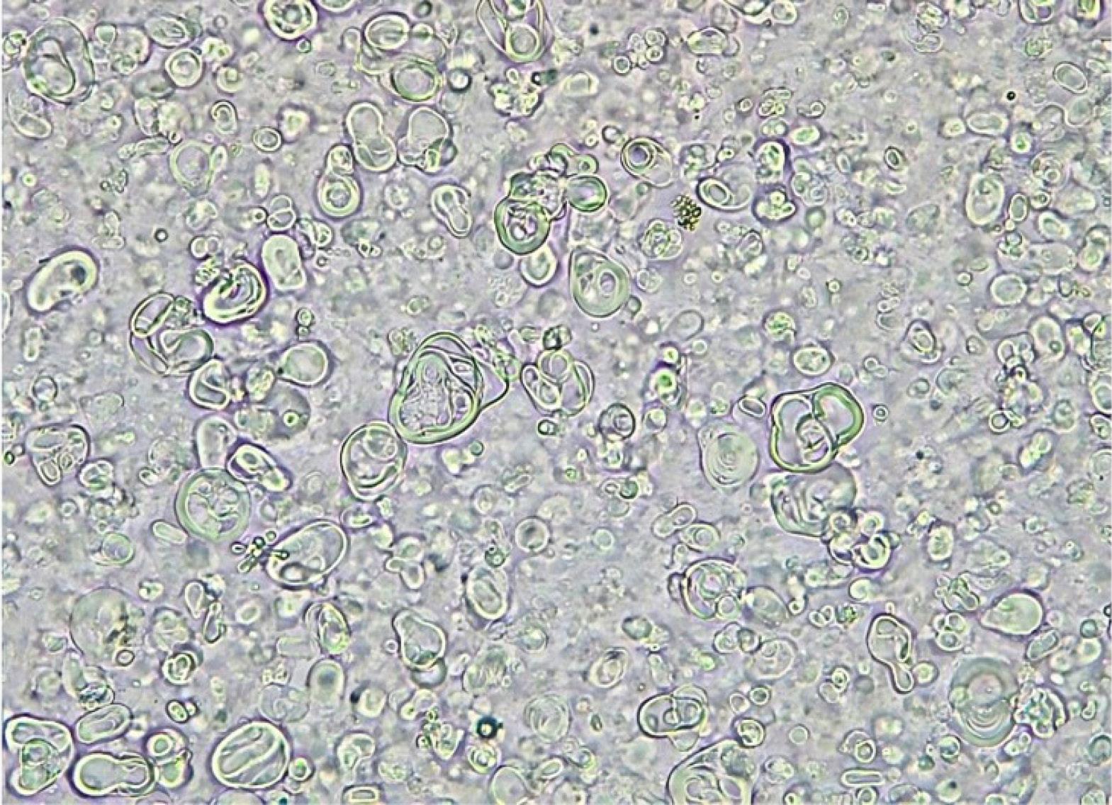
Figure 1.
Optical microscopy of YM niosomes composed of Brij 52/cholesterol 7:3 molar ratio with 10 × 40 magnification
.
Optical microscopy of YM niosomes composed of Brij 52/cholesterol 7:3 molar ratio with 10 × 40 magnification
Size Analysis and Physical Stability of YM Niosomes
As shown in Figure 2, the size distribution of YM niosomes exhibited a narrow pattern two weeks after preparation. The mean volume diameter (d v0.5) was 7.10 ± 0.20 µm for the YM niosomal suspension.
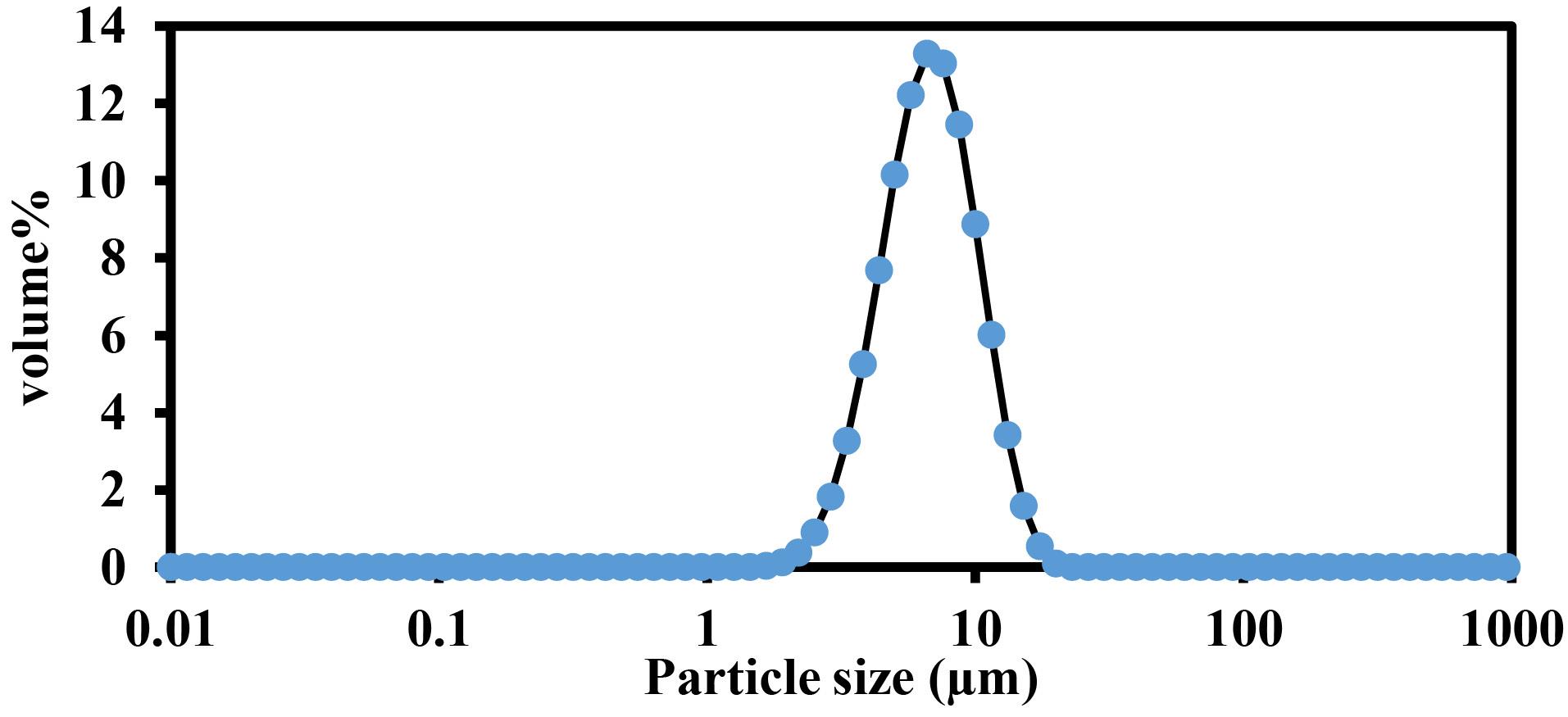
Figure 2.
Size distribution curve of Yerba mate niosomes, 2 weeks after preparation and storage at refrigerator temperature
.
Size distribution curve of Yerba mate niosomes, 2 weeks after preparation and storage at refrigerator temperature
Fluctuation of volume distribution of YM niosomes was measured by static laser scattering method during 6 months storage in refrigerator temperature (Figure 3). The size (dv0.5) of prepared niosomes was changed from 7.10 ± 0.20 to 7.36 ± 0.33 µm which indicates high physical stability of YM lipid vesicles.
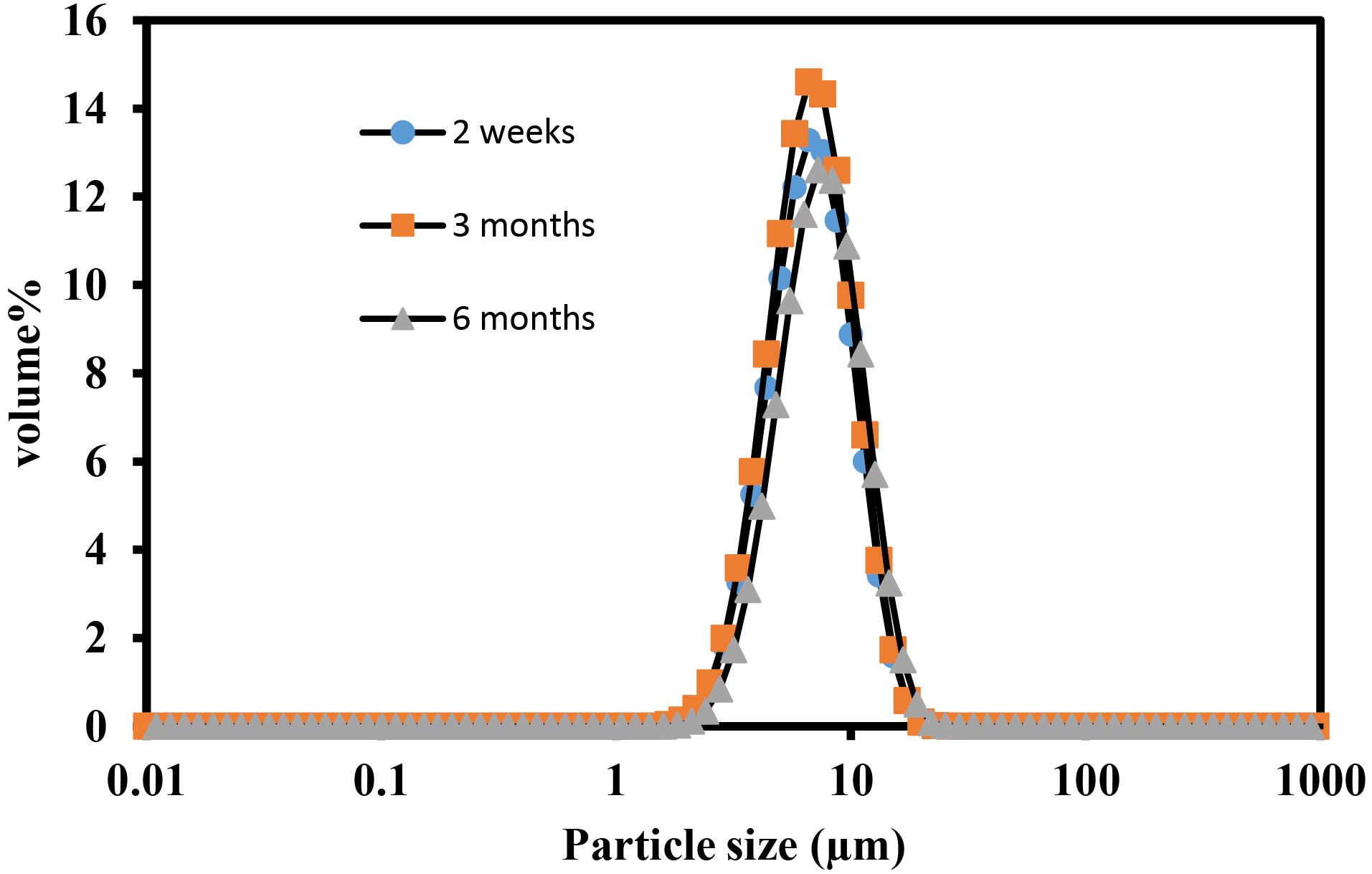
Figure 3.
Size distribution curves of YM niosomes during 6 months storage at refrigerator temperature as a description of niosomal suspension physical stability index
.
Size distribution curves of YM niosomes during 6 months storage at refrigerator temperature as a description of niosomal suspension physical stability index
Encapsulation Efficiency Percent of YM Extract in Niosomes
EE% of YM in Brij 52/cholesterol niosomes was measured as 63.25 ± 5.30 percent.
Physical Performance Measures
In the HIIT group, maximum running velocity increased progressively throughout the 8-week training period, with a statistically significant improvement observed during the later weeks (P < 0.0001; Figure 4A), while early changes within the first two weeks were not significant (P = 0.3149).
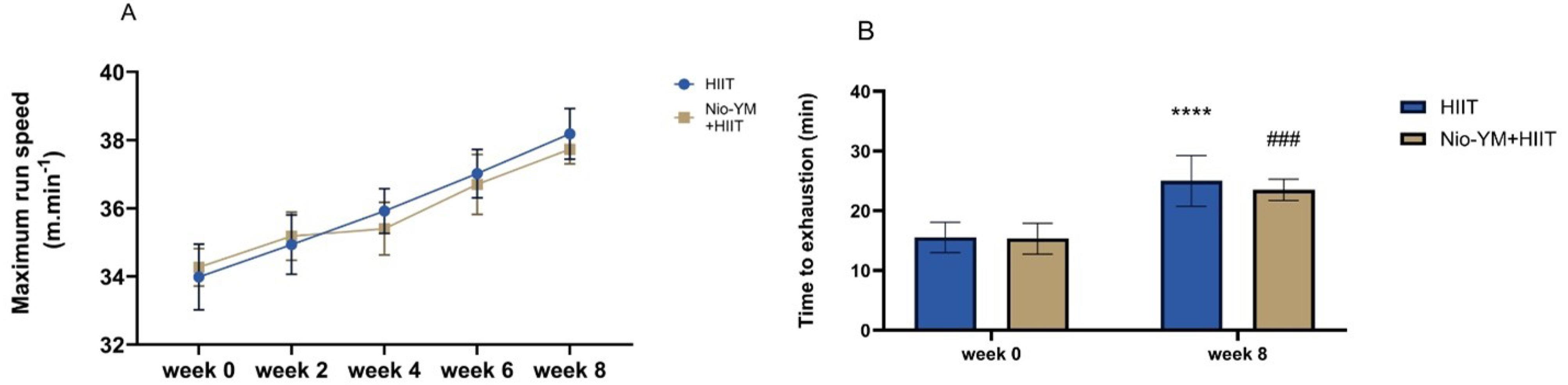
Figure 4.
Improvement of physical performance in rats subjected to (HIIT) and the combined intervention of niosomal Yerba Mate extract with HIIT. ****P < 0.0001 compared to the first week and ### P < 0.001 compared to the first week
.
Improvement of physical performance in rats subjected to (HIIT) and the combined intervention of niosomal Yerba Mate extract with HIIT. ****P < 0.0001 compared to the first week and ### P < 0.001 compared to the first week
In the Nio-YM + HIIT group, no significant improvement was detected up to week 4 compared with baseline (P = 0.3765); however, a pronounced increase was evident by the end of the intervention (P < 0.0001). Similarly, exercise tolerance significantly improved from baseline to week 8 in both the HIIT (P < 0.0001) and Nio-YM + HIIT (P = 0.0001) groups (Figure 4B), reflecting a marked enhancement in endurance capacity following both interventions. The total running distance, presented in Table 2, further supports the progressive improvement in physical performance across the intervention period.
Table 2.
Training volume program used in this study
|
Weeks
|
Distance (m)
|
Frequency (sessions/week)
|
Total Distance (m)
|
| Week 0-2 |
365 ( ± 2) |
3 |
1095 |
| Week 2-4 |
422 ( ± 2) |
3 |
1266 |
| Week 4-6 |
510 ( ± 1) |
3 |
1530 |
| Week 6-8 |
605 ( ± 3) |
3 |
1815 |
Cytokine and Oxidative Stress Measurement
Cytokine Measurement and Oxidative Stress Markers Statistical analysis revealed a significant elevation of IL-6 in the CS group compared to the CON group (P < 0.0001), confirming the pro-inflammatory impact of cigarette smoke exposure. Conversely, all intervention groups YM, Nio-YM, HIIT, and Nio-YM + HIIT exhibited significantly reduced IL-6 levels relative to the Veh group (P < 0.0001), highlighting their anti-inflammatory potential (Figure 5). However, no significant differences were observed among the interventions themselves.
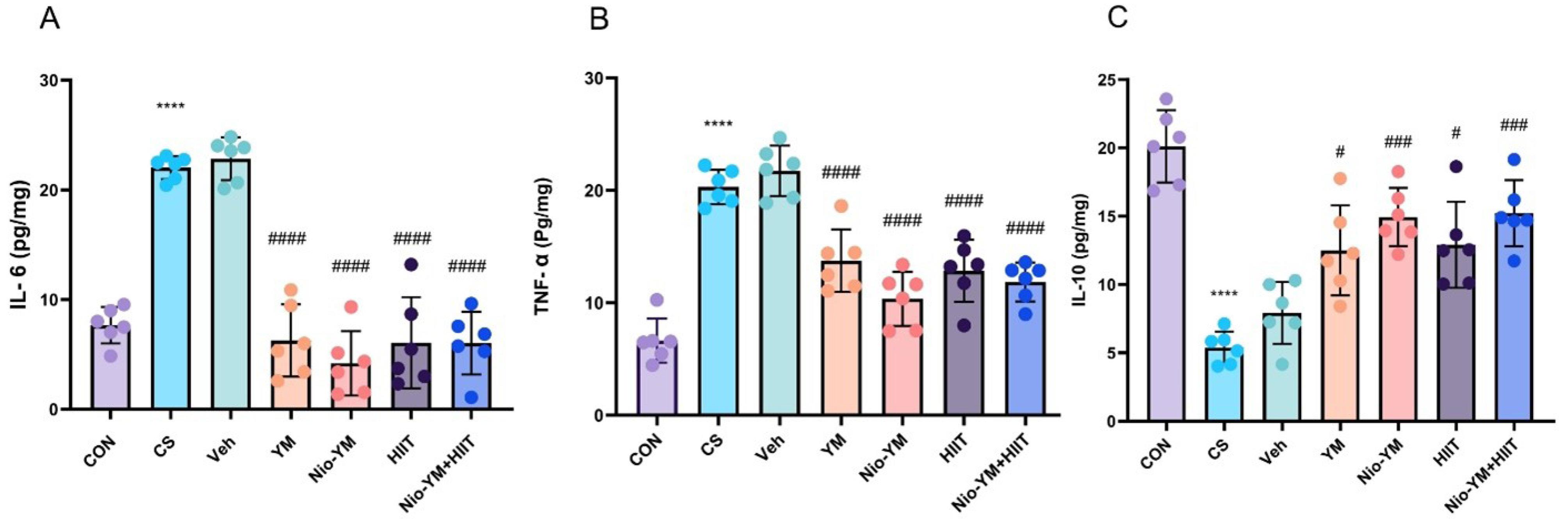
Figure 5.
Effect of HIIT, Nio-YM, their combination, and Yerba Mate extract alone on (A); IL-6, (B); TNF-α and (C); IL-10 in lung tissue. Experimental groups include Healthycontrol (CON), cigarette smoke (CS), vehicle control (Veh), yerba mate aqueous extract (YM), niosomal yerba mate aqueous extract (Nio-YM) and High intensity interval training (HIIT). ****P < 0.0001 compared to the CON group. Data are presented as mean ± SD for n = 6 in each group. ****P < 0.0001 compared to the CON group. # P < 0.05, ## P < 0.01, & #### P < 0.0001 compared to the Veh
.
Effect of HIIT, Nio-YM, their combination, and Yerba Mate extract alone on (A); IL-6, (B); TNF-α and (C); IL-10 in lung tissue. Experimental groups include Healthycontrol (CON), cigarette smoke (CS), vehicle control (Veh), yerba mate aqueous extract (YM), niosomal yerba mate aqueous extract (Nio-YM) and High intensity interval training (HIIT). ****P < 0.0001 compared to the CON group. Data are presented as mean ± SD for n = 6 in each group. ****P < 0.0001 compared to the CON group. # P < 0.05, ## P < 0.01, & #### P < 0.0001 compared to the Veh
Similarly, IL-10 concentrations were markedly suppressed in the CS group compared to CON (P < 0.0001), reflecting impaired anti-inflammatory signaling. Treatment with YM (P = 0.0494), Nio-YM (P = 0.0005), HIIT (P = 0.0248), and Nio-YM + HIIT (P = 0.0003) significantly restored IL-10 levels versus the Veh group, suggesting an enhancement of anti-inflammatory capacity.
Regarding TNF-α, the CS group showed a significant increase compared to CON (P < 0.0001), whereas all interventions led to a robust reduction (P < 0.0001 for all vs. Veh), underscoring their efficacy in attenuating smoke-induced inflammation.
In terms of oxidative stress markers (Figure 6), MDA levels were significantly elevated in the CS group (P < 0.0001), confirming increased lipid peroxidation. All treatment groups significantly lowered MDA levels relative to Veh (YM: P < 0.0001; Nio-YM: P = 0.0016; HIIT and Nio-YM + HIIT: P < 0.0001), indicating strong antioxidative effects.
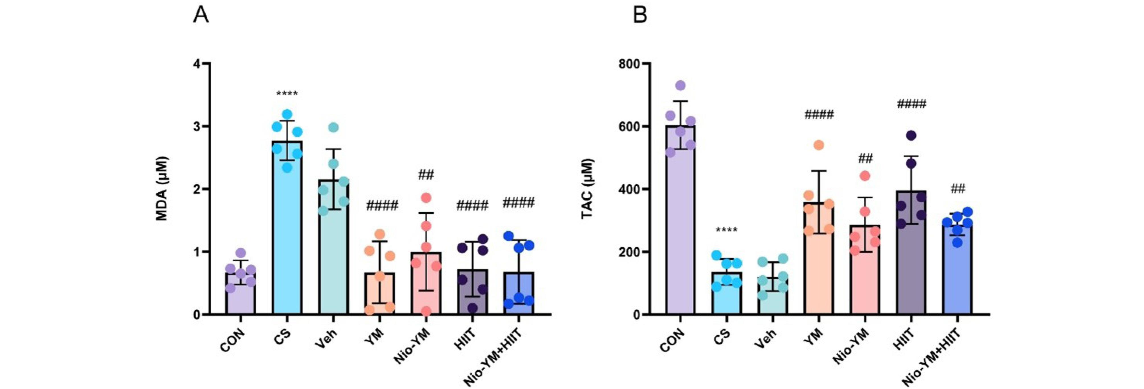
Figure 6.
Effect of HIIT, Nio-YM, their combination, and Yerba Mate extract alone on (A); MDA and (B); TAC in lung tissue. Healthycontrol (CON), cigarette smoke (CS), vehicle control (Veh), yerba mate aqueous extract (YM), niosomal yerba mate aqueous extract (Nio-YM) and High intensity interval training (HIIT). ****P < 0.0001 compared to the CON group. Data are presented as mean ± SD for n = 6 in each group. # P < 0.05, ## P < 0.01, & #### P < 0.0001 compared to the Veh
.
Effect of HIIT, Nio-YM, their combination, and Yerba Mate extract alone on (A); MDA and (B); TAC in lung tissue. Healthycontrol (CON), cigarette smoke (CS), vehicle control (Veh), yerba mate aqueous extract (YM), niosomal yerba mate aqueous extract (Nio-YM) and High intensity interval training (HIIT). ****P < 0.0001 compared to the CON group. Data are presented as mean ± SD for n = 6 in each group. # P < 0.05, ## P < 0.01, & #### P < 0.0001 compared to the Veh
TAC levels, which were markedly reduced in the CS group (P < 0.0001), significantly increased following all interventions (YM: P < 0.0001; Nio-YM: P = 0.0092; HIIT: P < 0.0001; Nio-YM + HIIT: P = 0.0086), reflecting restoration of antioxidant capacity. As with the cytokines, no statistically significant differences were found between the treatment groups.
Histopathology of Animal Lung Tissue
Pathological lung injury and airway epithelial damage scores were significantly elevated in the cigarette smoke and vehicle groups compared with controls (P = 0.0056). These groups also exhibited marked inflammatory cell infiltration and atelectasis (Figure 7). Among the intervention groups, only high-intensity interval training significantly attenuated the pathological alterations relative to the vehicle group (P = 0.0142), whereas Yerba Mate extract, niosomal Yerba Mate, and their combination failed to induce significant histopathological improvements.
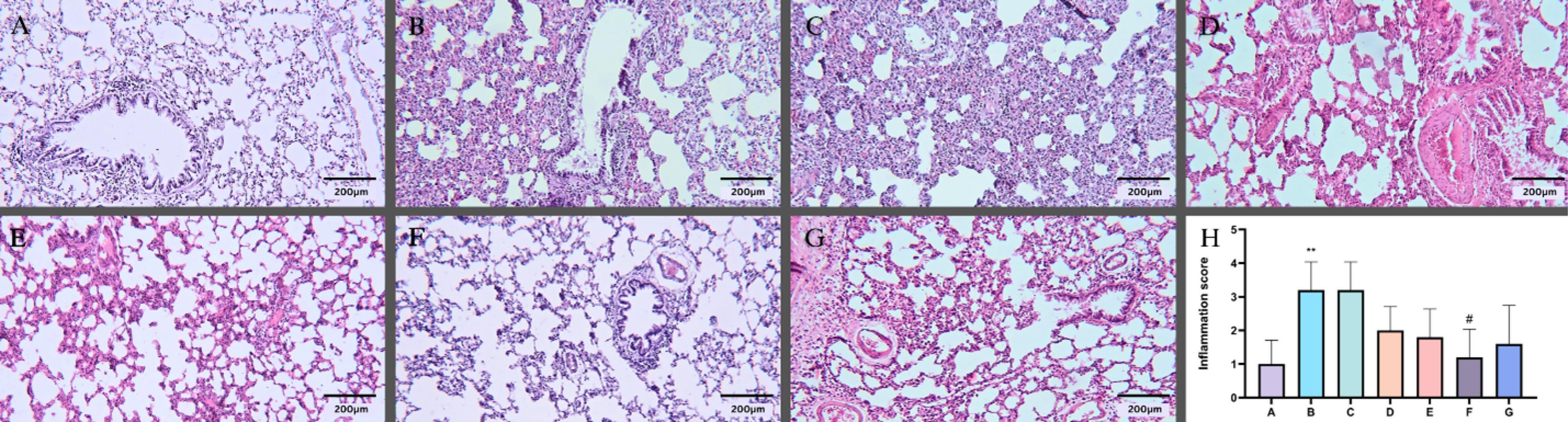
Figure 7.
Effect of HIIT, Nio-YM, their combination, and Yerba Mate extract alone on the microscopic presentation of cigarette smoke-induced acute lung injury in rats, as observed in hematoxylin and eosin-stained lung sections under light microscopy. A Healthycontrol group, B cigarette smoke group, C vehicle control group, D yerba mate aqueous extract group, E niosomal yerba mate aqueous extract group, F High intensity interval training group and G niosomal yerba mate aqueous extract + High intensity interval training group, H inflammation score in lung tissue. Data are presented as mean ± SD for n = 5 in each group. **P < 0.01 compared to the A group # P < 0.05 compared to the C
.
Effect of HIIT, Nio-YM, their combination, and Yerba Mate extract alone on the microscopic presentation of cigarette smoke-induced acute lung injury in rats, as observed in hematoxylin and eosin-stained lung sections under light microscopy. A Healthycontrol group, B cigarette smoke group, C vehicle control group, D yerba mate aqueous extract group, E niosomal yerba mate aqueous extract group, F High intensity interval training group and G niosomal yerba mate aqueous extract + High intensity interval training group, H inflammation score in lung tissue. Data are presented as mean ± SD for n = 5 in each group. **P < 0.01 compared to the A group # P < 0.05 compared to the C
Discussion
To the best of our knowledge, this is the first study to explore both the individual and combined effects of high-intensity interval training, Yerba Mate extract, and its niosomal formulation on systemic inflammation and oxidative stress in a rat model of cigarette smoke–induced lung injury. Non-ionic surfactants have long been utilized to improve bioavailability through various routes of administration, incorporated into vesicular and colloidal systems such as micelles (e.g., curcumin),34 microemulsions (e.g., enoxaparin),35 nano emulsions (e.g., isoformononetin)36 and niosomes, Nate glinide,37 carvedilol,38 quercetin,39 and Gingko biloba extract.40 Among these, polyoxyethylene alkyl ethers of the Brij® family have drawn particular attention. For instance, Brij 72 has been applied in oral paclitaxel delivery,41 while Brij 76 and Brij 78 have been incorporated as permeation enhancers in Span 60:cholesterol hybrid niosomes to improve oral absorption of 17-hydroxyprogesterone caproate.42 Similarly, Brij 52–cholesterol niosomes have shown improved pharmacokinetic profiles for nasal delivery of diltiazem in animal models.43 Consistent with our previous findings, where Brij 52-based lipid vesicles improved oral insulin delivery in diabetic rats,44 the current study employed the same surfactant to enhance the bioavailability of aqueous Yerba Mate extract.
Compared with the CS group, both YM–niosomes and YM–niosomes combined with HIIT elicited a significantly greater increase in IL-10 levels, reflecting a robust anti-inflammatory effect likely associated with the improved bioavailability and sustained release profile of the niosomal formulation.45
In addition to their biological efficacy, Brij 52-based niosomes demonstrated high physical stability, as shown in Figure 4. Particle size distribution analysis revealed minimal changes in vesicle diameter during six months of refrigerated storage, consistent with our earlier observations on Brij 52 niosomes for insulin delivery.46
The physicochemical characteristics of niosomes depend strongly on the preparation technique, which governs vesicle morphology and stability. Here, the thin-film hydration method was adopted, yielding multilamellar vesicles (MLVs). Upon oral administration, these vesicles undergo dynamic structural changes that facilitate the gradual release of encapsulated bioactive compounds into systemic circulation. In the small intestine, bile salts and pancreatic enzymes initially induce vesicle swelling followed by reduction in size.47 while enzymatic digestion of lipid bilayers results in controlled diffusion and erosion-driven release of the active ingredients.48 Furthermore, surfactants such as Triton X-100 and endogenous bile salts may integrate into lipid bilayers, transforming them into mixed micellar–bilayer structures.49 Nevertheless, the relatively large mean diameter of the prepared niosomes, together with the inherent complexity of intestinal absorption, may partly explain why pharmacokinetic differences between YM solution and YM–niosomes were not statistically significant in vivo.
High-intensity interval has emerged as a potent non-pharmacological strategy to regulate systemic and pulmonary immune responses in CS-induced lung injury and COPD.11,50,51 Chronic smoke exposure establishes a pro-inflammatory and oxidative milieu characterized by cytokine overproduction, immune cell infiltration, and oxidative damage to pulmonary tissue.52,53
Evidence from longitudinal studies indicates that regular moderate- to high-intensity exercise not only mitigates lung function decline but also reduces the risk of COPD development.54,55 In line with these findings, HIIT in the present study significantly elevated IL-10 and suppressed TNF-α compared with the CS group, confirming its potent anti-inflammatory potential. These effects are consistent with previous research demonstrating that exercise-induced myokines modulate immune signaling and attenuate systemic inflammation.56,57 The observed TNF-α reduction agrees with reports showing decreased airway inflammation following aerobic training,58,59 likely mediated through inhibition of STAT3 activation.60 However, other studies have reported no significant alterations in IL-6, TNF-α, or IL-10, indicating that the magnitude of the anti-inflammatory response may be contingent upon training intensity.61
Interestingly, IL-6 levels declined in our model, supporting its dual role as both pro- and anti-inflammatory mediator depending on physiological context.62-64 This pattern also aligns with epidemiological evidence linking greater physical activity to reduced systemic inflammation in smokers.65,66 Functionally, HIIT significantly enhanced time to exhaustion and maximal running speed in CS-exposed rats, supporting prior evidence that high-intensity interval training improves exercise tolerance and aerobic capacity.67,68
In accordance with a recent meta-analysis that emphasized a dose–response relationship between exercise intensity and improvements in pulmonary outcomes, the HIIT protocol applied in the present study was specifically structured to reflect these evidence-based thresholds, particularly within the optimal intensity ranges reported to enhance both functional capacity and respiratory performance.69
The antioxidant properties of Yerba Mate (YM) also contributed to the reduction of TNF-α levels, highlighting the close interplay between oxidative stress and inflammation. Consistent with previous findings, YM supplementation has been shown to lower TNF-α and IL-6 concentrations in diet-induced inflammatory models.70 Although its effects on acute lung injury remain insufficiently characterized, the present results suggest that YM may counteract cigarette smoke–induced oxidative damage. This interpretation is further supported by evidence demonstrating that YM attenuates lipid peroxidation (MDA) and strengthens antioxidant defenses via activation of the Nrf2 signaling pathway, which promotes glutathione synthesis.71-73 The elevation in TAC observed in both the HIIT and YM groups underscores their complementary roles in strengthening antioxidant capacity.74 Moreover, recent findings implicate the SOCS5/JAK2/STAT3 pathway in redox regulation,75 indicating that both HIIT and YM may target overlapping molecular mechanisms.
Importantly, relative to the CS group, a significant reduction in inflammatory histological scores was observed exclusively in the HIIT group. This improvement likely reflects adaptive remodeling of pulmonary structures driven by mechanical and metabolic stress during repeated high-intensity bouts.76
Collectively, these results suggest that HIIT primarily exerts its protective effects through biomechanical and metabolic pathways that foster tissue adaptation and systemic immunomodulation. In contrast, Yerba Mate acts predominantly through its polyphenolic antioxidants, suppressing lipid peroxidation and inflammatory cytokines. The niosomal formulation of YM further enhanced its efficacy, as reflected by the marked elevation of IL-10, likely due to improved bioavailability and sustained release of active compounds. However, combining HIIT with YM–niosomes did not yield additional synergistic benefits, possibly because both interventions modulate overlapping molecular pathways involved in inflammation and oxidative balance.
One limitation of this study was the absence of histological analyses of lung tissue, which could have provided structural evidence supporting the observed biochemical findings. Furthermore, the lack of lactometer measurements to quantify smoke intensity, along with the absence of total particulate matter assessment during cigarette smoke exposure, may have affected the precision of injury induction.
Conclusion
In summary, the present study demonstrated that both high-intensity interval training and niosome-encapsulated yerba mate extract effectively attenuated cigarette smoke–induced pulmonary injury in rats by modulating inflammatory cytokines and oxidative stress markers. Notably, their combination did not confer additive benefits. These findings suggest that HIIT may serve as a promising non-pharmacological approach for mitigating COPD-related lung damage.
Competing Interests
The authors state no conflict of interest.
Consent for Publication
Not applicable.
Data Availability Statement
All the data are included in this manuscript.
Ethical Approval
This study was conducted in accordance with ethical standards for animal research and approved by the relevant committee (IR.KMU.AEC.1404.046).
Acknowledgements
The authors would like to express their sincere gratitude to the Neuroscience Research Center of Kerman University of Medical Sciences for providing the research facilities and support. Special thanks are extended to Dr. Sheibani for his invaluable guidance and constructive contributions throughout the study.
References
- GBD 2019 Chronic Respiratory Diseases Collaborators. Global burden of chronic respiratory diseases and risk factors, 1990-2019: an update from the Global Burden of Disease Study 2019. EClinicalMedicine 2023; 59:101936. doi: 10.1016/j.eclinm.2023.101936 [Crossref] [ Google Scholar]
- Boers E, Barrett M, Su JG, Benjafield AV, Sinha S, Kaye L. Global burden of chronic obstructive pulmonary disease through 2050. JAMA Netw Open 2023; 6(12):e2346598. doi: 10.1001/jamanetworkopen.2023.46598 [Crossref] [ Google Scholar]
- Fabbri LM, Celli BR, Agustí A, Criner GJ, Dransfield MT, Divo M. COPD and multimorbidity: recognising and addressing a syndemic occurrence. Lancet Respir Med 2023; 11(11):1020-34. doi: 10.1016/s2213-2600(23)00261-8 [Crossref] [ Google Scholar]
- Vij N, Chandramani-Shivalingappa P, Van Westphal C, Hole R, Bodas M. Cigarette smoke-induced autophagy impairment accelerates lung aging, COPD-emphysema exacerbations and pathogenesis. Am J Physiol Cell Physiol 2018; 314(1):C73-87. doi: 10.1152/ajpcell.00110.2016 [Crossref] [ Google Scholar]
- Agustí A, Hogg JC. Update on the pathogenesis of chronic obstructive pulmonary disease. N Engl J Med 2019; 381(13):1248-56. doi: 10.1056/NEJMra1900475 [Crossref] [ Google Scholar]
- Faner R, Tal-Singer R, Riley JH, Celli B, Vestbo J, MacNee W. Lessons from ECLIPSE: a review of COPD biomarkers. Thorax 2014; 69(7):666-72. doi: 10.1136/thoraxjnl-2013-204778 [Crossref] [ Google Scholar]
- Bradford E, Jacobson S, Varasteh J, Comellas AP, Woodruff P, O’Neal W. The value of blood cytokines and chemokines in assessing COPD. Respir Res 2017; 18(1):180. doi: 10.1186/s12931-017-0662-2 [Crossref] [ Google Scholar]
- Rochester CL, Alison JA, Carlin B, Jenkins AR, Cox NS, Bauldoff G. Pulmonary rehabilitation for adults with chronic respiratory disease: an official American Thoracic Society clinical practice guideline. Am J Respir Crit Care Med 2023; 208(4):e7-26. doi: 10.1164/rccm.202306-1066ST [Crossref] [ Google Scholar]
- Ward TJ, Latimer L, Daynes E, Freeman SC, Ward S, Xu J. Impact of pulmonary rehabilitation programme design on effectiveness in COPD: a systematic review and component network meta-analysis. EClinicalMedicine 2025; 87:103433. doi: 10.1016/j.eclinm.2025.103433 [Crossref] [ Google Scholar]
- Toledo AC, Magalhaes RM, Hizume DC, Vieira RP, Biselli PJ, Moriya HT. Aerobic exercise attenuates pulmonary injury induced by exposure to cigarette smoke. Eur Respir J 2012; 39(2):254-64. doi: 10.1183/09031936.00003411 [Crossref] [ Google Scholar]
- Wang H, Liu Q, Liu L, Cao J, Liang Q, Zhang X. High-intensity interval training improves the outcomes of patients with chronic obstructive pulmonary disease: a meta-analysis of randomized controlled trials. Respir Med 2023; 208:107128. doi: 10.1016/j.rmed.2023.107128 [Crossref] [ Google Scholar]
- Qiao Z, Kou Z, Zhang J, Lv D, Cui X, Li D. Optimal intensity and type of lower limb aerobic training for patients with chronic obstructive pulmonary disease: a systematic review and network meta-analysis of RCTs. Ther Adv Respir Dis 2025; 19:17534666251323190. doi: 10.1177/17534666251323190 [Crossref] [ Google Scholar]
- José MF, Machado RP, Araujo PA, Speretta GF. Physiological effects of yerba maté (Ilex paraguariensis): a systematic review. Nutr Rev 2023; 81(9):1163-79. doi: 10.1093/nutrit/nuac109 [Crossref] [ Google Scholar]
- Song Y, Cao J, Cao F, Su E. A systematic review on the yerba mate (Ilex paraguariensis A St Hil). J Food Compost Anal 2025; 142:107466. doi: 10.1016/j.jfca.2025.107466 [Crossref] [ Google Scholar]
- Chen J, Li F, Li Z, McClements DJ, Xiao H. Encapsulation of carotenoids in emulsion-based delivery systems: enhancement of β-carotene water-dispersibility and chemical stability. Food Hydrocolloids 2017; 69:49-55. doi: 10.1016/j.foodhyd.2017.01.024 [Crossref] [ Google Scholar]
- Haddadzadegan S, Dorkoosh F, Bernkop-Schnürch A. Oral delivery of therapeutic peptides and proteins: technology landscape of lipid-based nanocarriers. Adv Drug Deliv Rev 2022; 182:114097. doi: 10.1016/j.addr.2021.114097 [Crossref] [ Google Scholar]
- Dumont C, Bourgeois S, Fessi H, Jannin V. Lipid-based nanosuspensions for oral delivery of peptides, a critical review. Int J Pharm 2018; 541(1-2):117-35. doi: 10.1016/j.ijpharm.2018.02.038 [Crossref] [ Google Scholar]
- Terzano C, Allegra L, Alhaique F, Marianecci C, Carafa M. Non-phospholipid vesicles for pulmonary glucocorticoid delivery. Eur J Pharm Biopharm 2005; 59(1):57-62. doi: 10.1016/j.ejpb.2004.06.010 [Crossref] [ Google Scholar]
- Akbarmehr A, Peighambardoust SH, Soltanzadeh M, Jafari SM, Sarabandi K. Microencapsulation of yerba mate extract: the efficacy of polysaccharide/protein hydrocolloids on physical, microstructural, functional, and antioxidant properties. Int J Biol Macromol 2023; 234:123678. doi: 10.1016/j.ijbiomac.2023.123678 [Crossref] [ Google Scholar]
- Scolaro B, Delwing-de Lima D, da Cruz JG, Delwing-Dal Magro D. Mate tea prevents oxidative stress in the blood and hippocampus of rats with acute or chronic ethanol administration. Oxid Med Cell Longev 2012; 2012:314758. doi: 10.1155/2012/314758 [Crossref] [ Google Scholar]
-
Kudo M, Gao M, Hayashi M, Kobayashi Y, Yang J, Liu T. Ilex paraguariensis A.St.-Hil. improves lipid metabolism in high-fat diet-fed obese rats and suppresses intracellular lipid accumulation in 3T3-L1 adipocytes via the AMPK-dependent and insulin signaling pathways. Food Nutr Res 2024;68. doi: 10.29219/fnr.v68.10307.
- Raeisi Estabragh MA, Behnam B, Torkzadeh-Mahani M, Pardakhty A. Niosome as a drug delivery carrier for sorafenib: preparation, investigation of physicochemical properties, and in vitro effects on HepG2 cell line. Adv Pharm Bull 2024; 14(4):836-45. doi: 10.34172/apb.43228 [Crossref] [ Google Scholar]
- Fatehi Chinar P, Bahraminejad S, Pardakhty A, Sharifi I, Ranjbar M, Karami-Mohajeri S. Preparation and evaluation of physicochemical properties and anti-leishmanial activity of zirconium/tioxolone niosomes against Leishmania major. Arab J Chem 2022; 15(10):104156. doi: 10.1016/j.arabjc.2022.104156 [Crossref] [ Google Scholar]
- Percie du Sert N, Hurst V, Ahluwalia A, Alam S, Avey MT, Baker M. The ARRIVE guidelines 20: updated guidelines for reporting animal research. J Cereb Blood Flow Metab 2020; 40(9):1769-77. doi: 10.1177/0271678x20943823 [Crossref] [ Google Scholar]
- Chen L, Sun BB, Wang T, Wang X, Li JQ, Wang HX. Cigarette smoke enhances β-defensin 2 expression in rat airways via nuclear factor-κB activation. Eur Respir J 2010; 36(3):638-45. doi: 10.1183/09031936.00029409 [Crossref] [ Google Scholar]
- Wang R, Tian H, Guo D, Tian Q, Yao T, Kong X. Impacts of exercise intervention on various diseases in rats. J Sport Health Sci 2020; 9(3):211-27. doi: 10.1016/j.jshs.2019.09.008 [Crossref] [ Google Scholar]
- Copp SW, Davis RT, Poole DC, Musch TI. Reproducibility of endurance capacity and VO2peak in male Sprague-Dawley rats. J Appl Physiol (1985) 2009; 106(4):1072-8. doi: 10.1152/japplphysiol.91566.2008 [Crossref] [ Google Scholar]
- Poole DC, Copp SW, Colburn TD, Craig JC, Allen DL, Sturek M. Guidelines for animal exercise and training protocols for cardiovascular studies. Am J Physiol Heart Circ Physiol 2020; 318(5):H1100-38. doi: 10.1152/ajpheart.00697.2019 [Crossref] [ Google Scholar]
- Bi P, Yue F, Sato Y, Wirbisky S, Liu W, Shan T. Stage-specific effects of Notch activation during skeletal myogenesis. Elife 2016; 5:e17355. doi: 10.7554/eLife.17355 [Crossref] [ Google Scholar]
- Kwak HB, Song W, Lawler JM. Exercise training attenuates age-induced elevation in Bax/Bcl-2 ratio, apoptosis, and remodeling in the rat heart. FASEB J 2006; 20(6):791-3. doi: 10.1096/fj.05-5116fje [Crossref] [ Google Scholar]
- Bejeshk MA, Beik A, Aminizadeh AH, Salimi F, Bagheri F, Sahebazzamani M. Perillyl alcohol (PA) mitigates inflammatory, oxidative, and histopathological consequences of allergic asthma in rats. Naunyn Schmiedebergs Arch Pharmacol 2023; 396(6):1235-45. doi: 10.1007/s00210-023-02398-5 [Crossref] [ Google Scholar]
- Bejeshk MA, Aminizadeh AH, Jafari E, Motamedi S, Zangiabadi I, Ghasemi A. Myrtenol ameliorates recognition memories’ impairment and anxiety-like behaviors induced by asthma by mitigating hippocampal inflammation and oxidative stress in rats. Neuroimmunomodulation 2023; 30(1):42-54. doi: 10.1159/000528626 [Crossref] [ Google Scholar]
- Ning Y, Shang Y, Huang H, Zhang J, Dong Y, Xu W. Attenuation of cigarette smoke-induced airway mucus production by hydrogen-rich saline in rats. PLoS One 2013; 8(12):e83429. doi: 10.1371/journal.pone.0083429 [Crossref] [ Google Scholar]
- Anjudikkal J, Shukla A, Pulikkal AK. Effects of micellar media on the bioavailability, solubility, and stability of curcumin. Next Nanotechnol 2025; 7:100124. doi: 10.1016/j.nxnano.2024.100124 [Crossref] [ Google Scholar]
- Alkrad JA, Assaf SM, Hussein-Al-Ali SH, Alrousan R. Microemulsions as nanocarriers for oral and transdermal administration of enoxaparin. J Drug Deliv Sci Technol 2022; 70:103248. doi: 10.1016/j.jddst.2022.103248 [Crossref] [ Google Scholar]
- Sultana N, Chauhan D, Yadav PK, Chourasia MK, Gayen JR. Enhanced oral bioavailability of isoformononetin through nanoemulsion: development, optimization, and characterization. J Pharm Innov 2024; 19(2):8. doi: 10.1007/s12247-024-09821-1 [Crossref] [ Google Scholar]
- Sultan AA, El-Gizawy SA, Osman MA, El Maghraby GM. Niosomes for oral delivery of nateglinide: in situ-in vivo correlation. J Liposome Res 2018; 28(3):209-17. doi: 10.1080/08982104.2017.1343835 [Crossref] [ Google Scholar]
- Arzani G, Haeri A, Daeihamed M, Bakhtiari-Kaboutaraki H, Dadashzadeh S. Niosomal carriers enhance oral bioavailability of carvedilol: effects of bile salt-enriched vesicles and carrier surface charge. Int J Nanomedicine 2015; 10:4797-813. doi: 10.2147/ijn.S84703 [Crossref] [ Google Scholar]
- Sadeghi-Ghadi Z, Ebrahimnejad P, Talebpour Amiri F, Nokhodchi A. Improved oral delivery of quercetin with hyaluronic acid containing niosomes as a promising formulation. J Drug Target 2021; 29(2):225-34. doi: 10.1080/1061186x.2020.1830408 [Crossref] [ Google Scholar]
- Jin Y, Wen J, Garg S, Liu D, Zhou Y, Teng L. Development of a novel niosomal system for oral delivery of Ginkgo biloba extract. Int J Nanomedicine 2013; 8:421-30. doi: 10.2147/ijn.S37984 [Crossref] [ Google Scholar]
- Bayindir ZS, Yuksel N. Characterization of niosomes prepared with various nonionic surfactants for paclitaxel oral delivery. J Pharm Sci 2010; 99(4):2049-60. doi: 10.1002/jps.21944 [Crossref] [ Google Scholar]
- Sharma PK, Kushwaha A, Repka MA, Murthy SN. Formulation development and pharmacokinetic investigation of self-assembled hybrid niosomes for oral delivery of 17-hydroxyprogesterone caproate. J Drug Deliv Sci Technol 2021; 61:102215. doi: 10.1016/j.jddst.2020.102215 [Crossref] [ Google Scholar]
- Ammar HO, Haider M, Ibrahim M, El Hoffy NM. In vitro and in vivo investigation for optimization of niosomal ability for sustainment and bioavailability enhancement of diltiazem after nasal administration. Drug Deliv 2017; 24(1):414-21. doi: 10.1080/10717544.2016.1259371 [Crossref] [ Google Scholar]
- Pardakhty A, Moazeni E, Varshosaz J, Hajhashemi V, Rouholamini Najafabadi A. Pharmacokinetic study of niosome-loaded insulin in diabetic rats. Daru 2011; 19(6):404-11. [ Google Scholar]
- Rajizadeh MA, Nematollahi MH, Jafari E, Bejeshk MA, Mehrabani M, Razeghinia MS. Niosome nanocarrier enhances the ameliorating effects of myrtenol in the lungs of rats with experimental asthma. OpenNano 2023; 11:100129. doi: 10.1016/j.onano.2023.100129 [Crossref] [ Google Scholar]
- Pardakhty A, Varshosaz J, Rouholamini A. In vitro study of polyoxyethylene alkyl ether niosomes for delivery of insulin. Int J Pharm 2007; 328(2):130-41. doi: 10.1016/j.ijpharm.2006.08.002 [Crossref] [ Google Scholar]
- Liu W, Ye A, Liu C, Liu W, Singh H. Structure and integrity of liposomes prepared from milk-or soybean-derived phospholipids during in vitro digestion. Food Res Int 2012; 48(2):499-506. doi: 10.1016/j.foodres.2012.04.017 [Crossref] [ Google Scholar]
- Liu W, Hou Y, Jin Y, Wang Y, Xu X, Han J. Research progress on liposomes: application in food, digestion behavior and absorption mechanism. Trends Food Sci Technol 2020; 104:177-89. doi: 10.1016/j.tifs.2020.08.012 [Crossref] [ Google Scholar]
- Nematollahi MH, Pardakhty A, Torkzadeh-Mahanai M, Mehrabani M, Asadikaram G. Changes in physical and chemical properties of niosome membrane induced by cholesterol: a promising approach for niosome bilayer intervention. RSC Adv 2017; 7(78):49463-72. doi: 10.1039/c7ra07834j [Crossref] [ Google Scholar]
- Patel S, Maddocks M, Man WD. Exercise training in COPD: FITT for purpose?. Chest 2020; 158(1):9-10. doi: 10.1016/j.chest.2020.02.040 [Crossref] [ Google Scholar]
-
Abbasi A, Wang D, Stringer WW, Casaburi R, Rossiter HB. Immune system benefits of pulmonary rehabilitation in chronic obstructive pulmonary disease. Exp Physiol. 2024. doi: 10.1113/ep091678.
- Barnes PJ. Inflammatory mechanisms in patients with chronic obstructive pulmonary disease. J Allergy Clin Immunol 2016; 138(1):16-27. doi: 10.1016/j.jaci.2016.05.011 [Crossref] [ Google Scholar]
- Tan DB, Fernandez S, Price P, French MA, Thompson PJ, Moodley YP. Impaired function of regulatory T-cells in patients with chronic obstructive pulmonary disease (COPD). Immunobiology 2014; 219(12):975-9. doi: 10.1016/j.imbio.2014.07.005 [Crossref] [ Google Scholar]
- Garvey C, Bayles MP, Hamm LF, Hill K, Holland A, Limberg TM. Pulmonary rehabilitation exercise prescription in chronic obstructive pulmonary disease: review of selected guidelines: an official statement from the American association of cardiovascular and pulmonary rehabilitation. J Cardiopulm Rehabil Prev 2016; 36(2):75-83. doi: 10.1097/hcr.0000000000000171 [Crossref] [ Google Scholar]
- Garcia-Aymerich J, Lange P, Benet M, Schnohr P, Antó JM. Regular physical activity modifies smoking-related lung function decline and reduces risk of chronic obstructive pulmonary disease: a population-based cohort study. Am J Respir Crit Care Med 2007; 175(5):458-63. doi: 10.1164/rccm.200607-896OC [Crossref] [ Google Scholar]
- Cavalcante PA, Gregnani MF, Henrique JS, Ornellas FH, Araújo RC. Aerobic but not resistance exercise can induce inflammatory pathways via toll-like 2 and 4: a systematic review. Sports Med Open 2017; 3(1):42. doi: 10.1186/s40798-017-0111-2 [Crossref] [ Google Scholar]
- Toledo-Arruda AC, Vieira RP, Guarnier FA, Suehiro CL, Caleman-Neto A, Olivo CR. Time-course effects of aerobic physical training in the prevention of cigarette smoke-induced COPD. J Appl Physiol (1985) 2017; 123(3):674-83. doi: 10.1152/japplphysiol.00819.2016 [Crossref] [ Google Scholar]
- Vieira RP, Claudino RC, Duarte AC, Santos AB, Perini A, Faria Neto HC. Aerobic exercise decreases chronic allergic lung inflammation and airway remodeling in mice. Am J Respir Crit Care Med 2007; 176(9):871-7. doi: 10.1164/rccm.200610-1567OC [Crossref] [ Google Scholar]
- Chen HI, Hsieh SY, Yang FL, Hsu YH, Lin CC. Exercise training attenuates septic responses in conscious rats. Med Sci Sports Exerc 2007; 39(3):435-42. doi: 10.1249/mss.0b013e31802d11c8 [Crossref] [ Google Scholar]
- Wang X, Wang Z, Tang D. Aerobic exercise alleviates inflammation, oxidative stress, and apoptosis in mice with chronic obstructive pulmonary disease. Int J Chron Obstruct Pulmon Dis 2021; 16:1369-79. doi: 10.2147/copd.S309041 [Crossref] [ Google Scholar]
- Wang CH, Chou PC, Joa WC, Chen LF, Sheng TF, Ho SC. Mobile-phone-based home exercise training program decreases systemic inflammation in COPD: a pilot study. BMC Pulm Med 2014; 14:142. doi: 10.1186/1471-2466-14-142 [Crossref] [ Google Scholar]
- Muñoz-Cánoves P, Scheele C, Pedersen BK, Serrano AL. Interleukin-6 myokine signaling in skeletal muscle: a double-edged sword?. FEBS J 2013; 280(17):4131-48. doi: 10.1111/febs.12338 [Crossref] [ Google Scholar]
- Peake JM, Della Gatta P, Suzuki K, Nieman DC. Cytokine expression and secretion by skeletal muscle cells: regulatory mechanisms and exercise effects. Exerc Immunol Rev 2015; 21:8-25. [ Google Scholar]
- van der Vlist J, Janssen TW. The potential anti-inflammatory effect of exercise in chronic obstructive pulmonary disease. Respiration 2010; 79(2):160-74. doi: 10.1159/000233264 [Crossref] [ Google Scholar]
- Loprinzi PD, Walker JF, Lee H. Association between physical activity and inflammatory markers among US adults with chronic obstructive pulmonary disease. Am J Health Promot 2014; 29(2):81-8. doi: 10.4278/ajhp.130510-QUAN-235 [Crossref] [ Google Scholar]
- Rajizadeh MA, Haj Hosseini M, Bahrami M, Bahri F, Rostamabadi F, Bagheri F. High-intensity intermittent training ameliorates methotrexate-induced acute lung injury. BMC Pulm Med 2024; 24(1):45. doi: 10.1186/s12890-024-02853-w [Crossref] [ Google Scholar]
- Seldeen KL, Lasky G, Leiker MM, Pang M, Personius KE, Troen BR. High intensity interval training improves physical performance and frailty in aged mice. J Gerontol A Biol Sci Med Sci 2018; 73(4):429-37. doi: 10.1093/gerona/glx120 [Crossref] [ Google Scholar]
- Li FH, Sun L, Zhu M, Li T, Gao HE, Wu DS. Beneficial alterations in body composition, physical performance, oxidative stress, inflammatory markers, and adipocytokines induced by long-term high-intensity interval training in an aged rat model. Exp Gerontol 2018; 113:150-62. doi: 10.1016/j.exger.2018.10.006 [Crossref] [ Google Scholar]
- Xie J, Guo J, Wang B. Optimal exercise modalities and doses for alleviating dyspnea symptoms and enhancing exercise capacity in patients with chronic obstructive pulmonary disease: a network and dose-response meta-analysis. Arch Phys Med Rehabil 2025; 106(10):1594-602. doi: 10.1016/j.apmr.2025.05.001 [Crossref] [ Google Scholar]
- Arçari DP, Bartchewsky W, dos Santos TW, Oliveira KA, Funck A, Pedrazzoli J. Antiobesity effects of yerba maté extract (Ilex paraguariensis) in high-fat diet-induced obese mice. Obesity (Silver Spring) 2009; 17(12):2127-33. doi: 10.1038/oby.2009.158 [Crossref] [ Google Scholar]
- Camara FD, Pedroso GS, Roman SS, Dallago RM, Valduga AT, Fernandes BB. Yerba mate (Ilex paraguariensis St Hil) extract inhibits hand-rolled cornhusk cigarette smoke-induced oxidative pulmonary damage. An Acad Bras Cienc 2020; 92(2):e20191141. doi: 10.1590/0001-3765202020191141 [Crossref] [ Google Scholar]
- Gao H, Liu Z, Wan W, Qu X, Chen M. Aqueous extract of yerba mate tea lowers atherosclerotic risk factors in a rat hyperlipidemia model. Phytother Res 2013; 27(8):1225-31. doi: 10.1002/ptr.4856 [Crossref] [ Google Scholar]
- Moskaug J, Carlsen H, Myhrstad MC, Blomhoff R. Polyphenols and glutathione synthesis regulation. Am J Clin Nutr 2005; 81(1 Suppl):277S-83S. doi: 10.1093/ajcn/81.1.277S [Crossref] [ Google Scholar]
- Puangpraphant S, de Mejia EG. Saponins in yerba mate tea (Ilex paraguariensis A St-Hil) and quercetin synergistically inhibit iNOS and COX-2 in lipopolysaccharide-induced macrophages through NFκB pathways. J Agric Food Chem 2009; 57(19):8873-83. doi: 10.1021/jf902255h [Crossref] [ Google Scholar]
- Lei Y, He J, Hu F, Zhu H, Gu J, Tang L. Sequential inspiratory muscle exercise-noninvasive positive pressure ventilation alleviates oxidative stress in COPD by mediating SOCS5/JAK2/STAT3 pathway. BMC Pulm Med 2023; 23(1):385. doi: 10.1186/s12890-023-02656-5 [Crossref] [ Google Scholar]
- Nymand SB, Hartmann JP, Ryrsø CK, Rossen NS, Christensen RH, Iepsen UW. Exercise adaptations in COPD: the pulmonary perspective. Am J Physiol Lung Cell Mol Physiol 2022; 323(6):L659-66. doi: 10.1152/ajplung.00549.2020 [Crossref] [ Google Scholar]