Pharmaceutical Sciences. 31(4):491-496.
doi: 10.34172/PS.025.42969
Research Article
Assessment of the Protective Effects of Omega-3 Fatty acid and Vitamin E Against Carbamazepine Hepatotoxicity in Rats by Alteration of Liver Enzymes, Antioxidant and Inflammatory Markers
Furkan Majid Nassir AL Attar Conceptualization, Formal analysis, Investigation, Writing – original draft, 1, * 
Intesar Tarik Numan Formal analysis, Writing – review & editing, 2
Suhad Faisal Hatem Al-Mugdadi Conceptualization, Formal analysis, Writing – review & editing, 3
Author information:
1Department of Pharmacology and Toxicology, College of Pharmacy, Mustansiriyah University, Baghdad, Iraq
2Department of Pharmacy, Alnukhba University College, Baghdad, Iraq
3Department of Clinical Laboratories Sciences, College of Pharmacy, Mustansiriyah University, Baghdad, Iraq
Abstract
Background:
Drug-induced liver injury (DILI) is a significant public health problem. Carbamazepine (CBZ), an antiepileptic medicine used in the treatment of epilepsy, has been associated with the development of DILI, potentially progressing to liver failure. Previous studies suggested that omega-3 fatty acids and vitamin E possess antioxidant and anti-inflammatory characteristics. However, the protective roles of omega-3 fatty acids and vitamin E against CBZ-induced toxicity remain controversial.
Methods:
Thirty rats were randomly divided into five groups: group I (control), group II, (CBZ only), group III (CBZ+omega-3 fatty acids), group IV (CBZ+vitamin E), and group V (CBZ+omega-3 fatty acids and vitamin E). Liver enzymes, oxidative stress biomarkers (glutathione, malondialdehyde), proinflammatory cytokines, and cytokeratin 18 gene expression levels were assessed using standard techniques.
Results:
Liver enzymes, proinflammatory cytokines, malondialdehyde and cytokeratin 18 gene expressions reduced significantly (P<0.01) in groups III, IV, and V in comparison to group II, while glutathione was higher than in the induction group II (P<0.01).
Conclusion:
These findings suggest that omega-3 fatty acids and vitamin E exert protective effects against CBZ-induced hepatotoxicity, likely through antioxidant and anti-inflammatory mechanisms.
Keywords: Carbamazepine, Drug-induced liver injury, Liver, Omega-3 fatty acid, Vitamin E
Copyright and License Information
© 2025 The Author(s).
This is an open access article and applies the Creative Commons Attribution Non-Commercial License (
http://creativecommons.org/licenses/by-nc/4.0/). Non-commercial uses of the work are permitted, provided the original work is properly cited.
Funding Statement
The authors stated that no funding was provided.
Introduction
The liver is a large gland and the second largest organ in the body, weighing up to 1500 g. It is responsible for metabolism, and detoxification processes.1 Drug-induced liver injury (DILI) is a severe complication and serious adverse effect caused by a wide range of drugs.2 DILI management typically consists of immediate discontinuation of the treatment and early therapeutic intervention to prevent severe complications.3
Carbamazepine (CBZ) was first discovered and synthesized by the Swiss chemist Walter Schindler in 1953. It is a white crystalline powder that is poorly soluble in water. Due to its lipophilic property, it crosses the blood brain barrier and affects the brain.4 It is widely prescribed for various medical conditions including epilepsy, neuropathic pain, schizophrenia, and bipolar disorder. CBZ is metabolized via oxidation mediated by enzymes like CYP3A4 and CYP2C8, which leads to the formation of CBZ-10,11-epoxide, an active metabolite associated with toxic effects. Its metabolic pathway includes hydroxylation of the 6-membered aromatic rings and N-glucuronidation of the carbamoyl side chain.5
CBZ acute intoxication at high doses is clinically rare. However, CBZ intoxication can occur in combination with other kinds of substances like benzodiazepines or alcohol.6 CBZ overdose can affect many systems, and the toxicity can range from mild to severe. Its intoxication presents with many clinical features such as neurotoxicity, ataxia, altered consciousness and coma. Cardiovascular toxicity includes tachycardia, myocardial depression, atrioventricular block, and severe hepatitis.7
Omega-3 fatty acids are found in a variety of food sources, and they are involved in regulating blood clotting and inflammation.8 They are typically absorbed in the human intestines and incorporated into cell membranes. Omega-3 fatty acids exhibit anti-inflammatory and antioxidant effects, reducing inflammatory responses. They help prevent cardiovascular conditions, reduces hypertension, and improves blood circulation and also possesses protective properties for the heart and kidneys through modification of the antioxidant, inflammatory, and apoptosis biomarkers.9,10 Omega-3 fatty acids have a preventive effect against liver disease.11
Vitamin E is a lipid-soluble substance with multiple functions, such as antioxidants, anti-inflammatory, and anticancer activities.12 Vitamin E deficiency causes neuromuscular impairments and inflammation.13
Previous studies have demonstrated that omega-3 fatty acids provide considerable protective effects against hepatotoxicity caused by various medications like methotrexate,14 doxorubicin,15 lead acetate,16 and mercury chloride.17 Vitamin E has also been shown to protect against liver damage induced by medications like doxorubicin15 and imidacloprid,18 both alone and in combination with omega-3. Nevertheless, the potential advantages of their individual or combined administration against CBZ-induced hepatotoxicity remain unexplored. According to their antioxidant and anti-inflammatory properties, it is possible that the two medications will give better protection when provided together, compared to each supplement individually, against CBZ-induced hepatotoxicity. The aim of this study is to investigate the potential protective effects of omega-3 fatty acids and vitamin E on CBZ-induced hepatotoxicity in rats.
Methods
Materials
CBZ was obtained from Novartis (Switzerland) in suspension form containing 2%. Omega-3 fatty acids including eicosapentaenoic acid (EPA) and docosahexaenoic acid (DHA), at a dose of 400 mg, were sourced from Solgar (United States) and vitamin E (alpha-tocopherol) was purchased from Meryer (China). Ketamine (10%) and Xylazine (2%) were both purchased from Alfasan (Woerden, Netherlands).
Animals
Adult male albino Wistar rats weighing 180-200 g and aged 16–18 weeks were used. The animals were properly housed in cages in the animal facility of College of Pharmacy, Mustansiriyah University, under controlled temperature (25 ℃) and humidity 50-55% conditions.
Experimental Design
Thirty rats were divided into five groups, with six rats in each group:
-
Group 1 (Control): received distilled water alone, orally once daily for 14 days.
-
Group 2 (CBZ): Treated with CBZ (100 mg/kg/day, orally) for 14 days.
-
Group 3 (CBZ + omega-3): Treated with CBZ (100 mg/kg/day, orally) then followed by omega-3 including EPA + DHA (400 mg/kg/day, orally) one hour later, for 14 days.
-
Group 4 (CBZ + vitamin E): Treated with CBZ (100 mg/kg/day, orally), followed by vitamin E (400 IU/kg/day, orally) one hour later, for 14 days.
-
Group 5 (CBZ + omega-3 fatty acids + vitamin E): Treated with CBZ (100 mg/kg/day, orally), followed by a combination of omega-3 fatty acids including EPA + DHA (400 mg/kg/day) and vitamin E (400 IU/kg/day) one hour later, for 14 days.
After two weeks, the rats were anesthetized by 0.2 mL xylazine 2% and 0.1 mL ketamine 10%.
Blood Sample Collection and Liver Dissection
Three mL of blood were collected via cardiac puncture, allowed to clot at room temperature for 30 minutes, then centrifugated at 3000 rpm for 15 minutes. The serum was then stored at -80°C. The liver was dissected; the first portion homogenized for analysis of malondialdehyde (MDA) and glutathione (GSH), while the second portion was stored in Trizol for analysis of cytokeratin 18 (CK-18) gene expression.
Biochemical Analysis
Biochemical analysis, serum samples were used to analyze liver function markers. Liver enzymes are crucial indicators for impaired liver function, and their marked elevation indicates severe liver injury. To assess liver enzyme activity, alanine aminotransferase (ALT), aspartate aminotransferase (AST), and alkaline phosphatase (ALP) levels were measured according to the company’s protocols (Linear Chemicals, Spain).
Assessment of Oxidative Stress Biomarkers
The levels of oxidative stress biomarkers i.e., GSH and MDA were measured using MDA ELISA Kit (E-EL- 0060, USA), and GSH ELISA Kit (E-EL-0026, USA). These markers of oxidative stress are considered hallmarks for hepatotoxicity due to their presence in liver tissue.
Assessment of Inflammatory Parameters
The values of tumor necrosis factor-alpha (TNF-α) and interleukin 6 (IL-6) were quantified using ELISA. These inflammatory markers are pro-inflammatory cytokines commonly used to evaluate the toxic effect of drugs. Detection of these biomarkers was performed according to the manufacturer’s instructions (TNF- alpha kit: SEA133Ra, China; IL-6 kit: SEA079Ra, China).
Molecular Approach
CK-18 gene expression is commonly used as a hallmark of drug-induced hepatotoxicity.19 CK-18 gene expression measured by using real-time (RT-PCR). For RNA extraction, TranZol Up Plus RNA reagent (China) was used. The extracted RNA was then used to synthesize complementary DNA (cDNA) using the EasyScript® First-Strand cDNA Synthesis SuperMix kit (Transgen, China). All primers were lyophilized and included both target and endogenous genes (HKG2), Primer sequence (5’-3’) were as follows:
Working primer concentration was prepared at 10 pmol/µL for the qRT –PCR reaction mixture. The final reaction volume was 25 µL, containing: 8 µL cDNA, 1 µL each of forward and reverse Primers, 8 µL of qPCR Master Mix (SYBR green), and 7 µL of double-distilled water (ddH2O).
Amplification was performed by incubations of mixture to SaCycler_96 Real-Time PCR system under following conditions:
-
Enzyme activation: 94 °C for 30 seconds (for one cycle)
-
Denaturation: 94 °C for 5 seconds
-
Annealing and extension: 60 °C for 35 seconds (for 45 cycles)
-
High-resolution melting: 90 °C for 15 seconds (for 100 cycles)
The expression of the target gene levels was assessed using the relative quantitative method for all groups, calculated as: Fold change = 2-∆∆Ct
Statistical Analysis
The statistical analysis was performed using the Statistical Package for the Social Sciences (SPSS), version 2019. One-way ANOVA followed by the LSD post hoc test was used to compare significant differences between group means in the study. A P value of < 0.05 was considered statistically significant, while P < 0.01 was considered highly significant.
Results
Liver Function
ALT levels in group 2 (CBZ) (33.50 ± 2.54 U/L) were significantly higher compared to group 1 (control) (27.90 ± 1.36 U/L). When treated with omega-3 fatty acids, the mean serum ALT level was lower than group 2, but without a significant difference (28.40 ± 1.63 U/L). Moreover, serum ALT levels were significantly reduced in groups treated with vitamin E alone and the combination of vitamin E and omega-3 fatty acids (27.20 ± 1.17 U/L and 26.00 ± 1.04 U/L, respectively), with P < 0.05 compared to group 2.
The mean serum level of AST was elevated in group 2 (70.00 ± 3.51 U/L) compared to group 1 (42.01 ± 2.47 U/L). There was a highly significant reduction in the AST levels in groups 3, 4, and 5, treated with omega-3 fatty acids, vitamin E, and their combination (47.90 ± 2.94 U/L,56.50 ± 3.05 U/L, 45.15 ± 2.37 U/L, respectively), with P < 0.001.
The mean serum ALP level in the induction group with CBZ (group 2) was significantly higher (48.17 ± 2.74 U/L) than in the control group (33.08 ± 2.19 U/L).While the treated groups with omega-3 and vitamin E (groups 3, 4 and 5) showed a highly significant reduction in ALP levels (33.95 ± 2.07 U/L, 30.40 ± 1.69 U/L, and 29.48 ± 1.54 U/L, respectively), P < 0.01 versus the induction group (group 2). The results are detailed in Table 1.
Table 1.
Effect of omega- 3 fatty acids and vitamin E on ALT, AST and ALP liver enzyme levels in carbamazepine-induced hepatotoxicity in rats
|
Group
|
ALT Mean±SE (U/L)
|
AST Mean±SE (U/L)
|
ALP Mean±SE (IU/L)
|
| Group 1 |
27.90 ± 1.36b |
42.01 ± 2.47c |
33.08 ± 2.19b |
| Group 2 |
33.50 ± 2.54a |
70.00 ± 3.51a |
48.17 ± 2.74a |
| Group 3 |
28.40 ± 1.63ab |
47.90 ± 2.94c |
33.95 ± 2.07b |
| Group 4 |
27.20 ± 1.17b |
56.50 ± 3.05b |
30.40 ± 1.69b |
| Group 5 |
26.00 ± 1.04b |
45.15 ± 2.37c |
29.48 ± 1.54b |
Data were expressed as mean ± SE, mean superscript with different lowercase letters (a, b, c, d) differ statistically, LSD used to compare mean among groups, P value ≤ 0.05 * (significant difference), P-value ≤ 0.01 ** (highly significant difference).
Oxidative Stress Markers
The mean of GSH level in homogenized liver tissue of group 2 was significantly lower (5.52 ± 0.37 U/L) compared to group 1 (14.08 ± 0.64 U/L). Treatment with omega-3 (group 3) showed no significant differences from group 2. However, GSH levels were significantly increased in the groups treated with vitamin E alone (16.016 ± 0.89 U/L) and the combination of omega-3 and vitamin E group (19.345 ± 1.03 U/L), compared to group 2 (Figure 1).
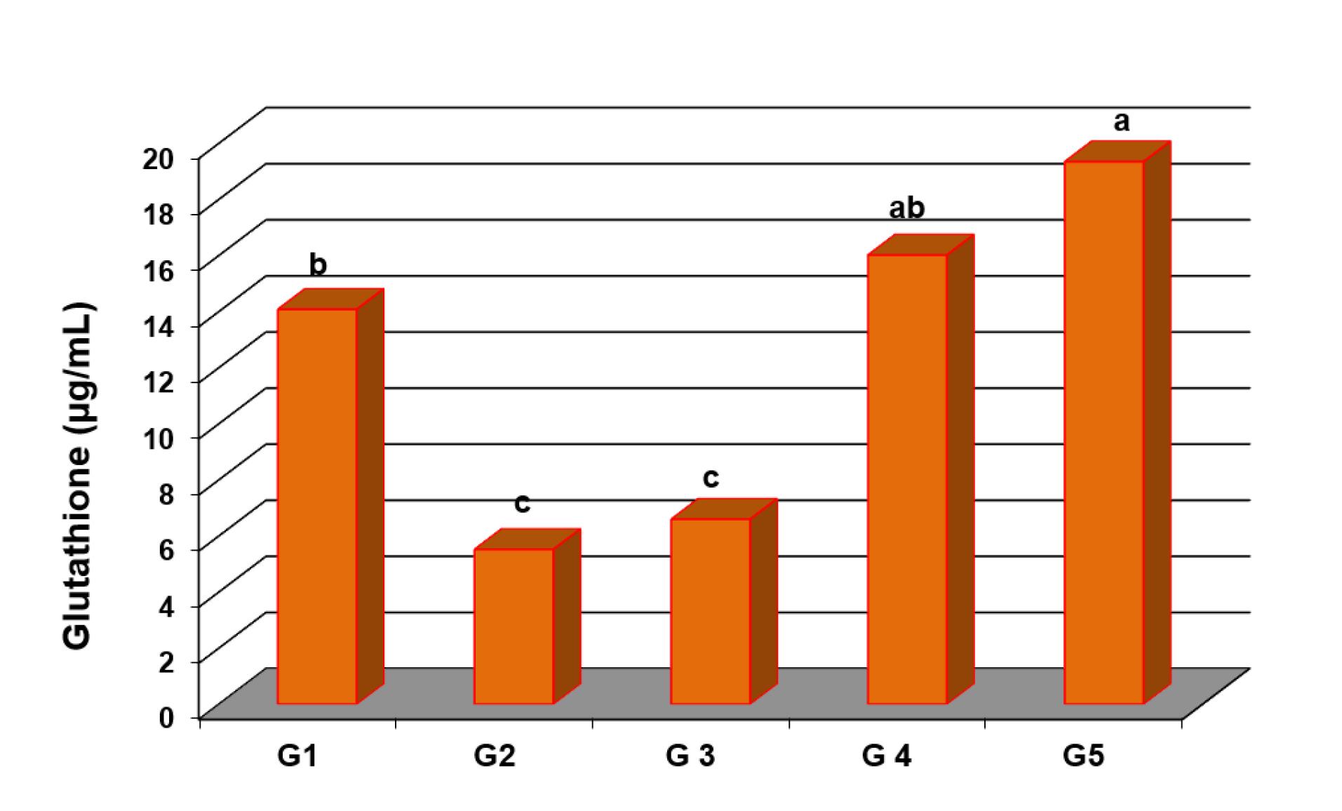
Figure 1.
Comparison between different groups regarding GSH levels. G1: Control group, G2: induction group with CBZ, G3: omega-3 fatty acids with induction, G4: Vitamin E with induction, and G5: Combination of omega-3 fatty acids and vitamin E with induction. Different letters (a, b and c) indicate statistically significant differences
.
Comparison between different groups regarding GSH levels. G1: Control group, G2: induction group with CBZ, G3: omega-3 fatty acids with induction, G4: Vitamin E with induction, and G5: Combination of omega-3 fatty acids and vitamin E with induction. Different letters (a, b and c) indicate statistically significant differences
MDA levels in homogenized liver tissue were significantly higher in group 2 (232.38 ± 11.37 U/L) compared to group 1 (120.98 ± 7.04 U/L). Treated groups (group 3, 4 and 5) showed a significant reduction in MDA levels (222.17 ± 8.75 U/L, 172.62 ± 8.62 U/L, 138.45 ± 6.59 U/L, respectively), with P < 0.01 compared to group 2 (Figure 2).
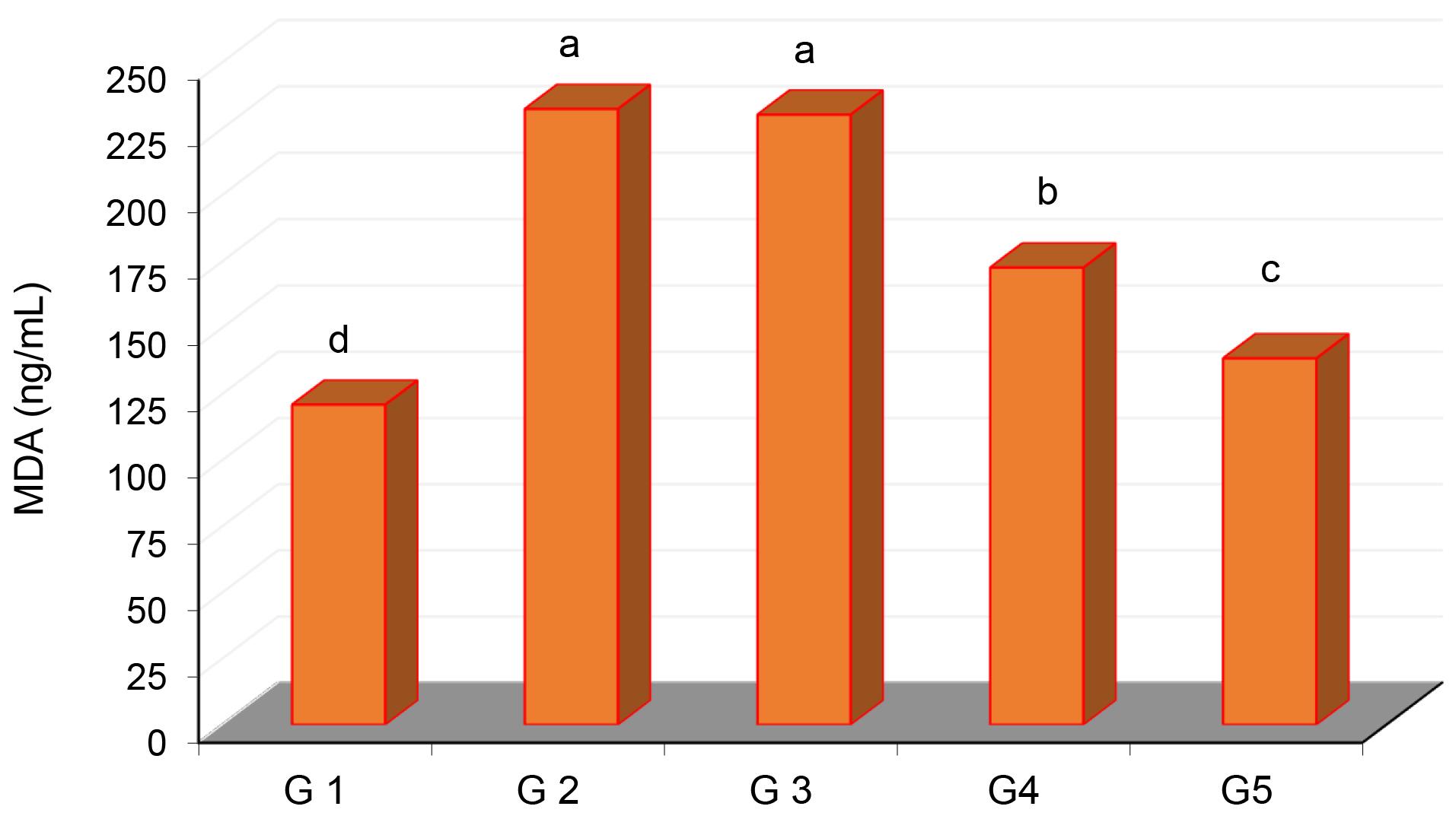
Figure 2.
Comparison between different groups regarding MDA levels. G1: Control group, G2: induction group with CBZ, G3: omega-3 fatty acids with induction, G4: vitamin E with induction, and G5: combination of omega-3 fatty acids and vitamin E with induction. Different letters (a, b and c) indicate statistically significant differences
.
Comparison between different groups regarding MDA levels. G1: Control group, G2: induction group with CBZ, G3: omega-3 fatty acids with induction, G4: vitamin E with induction, and G5: combination of omega-3 fatty acids and vitamin E with induction. Different letters (a, b and c) indicate statistically significant differences
Inflammatory Cytokines Markers
The mean serum TNF-α level in group 2 was significantly high (391.807 ± 23.44 U/L) than in the control group (269.336 ± 15.02 U/L). While treated groups (group 3, 4 and 5) showed significant reductions (340.416 ± 19.52 U/L, 335.565 ± 16.87 U/L, and 209.533 ± 12.63 U/L, respectively), with P ≤ 0.01 compared to group 2. Group 5 showed the greatest reduction in serum TNF-α level. (Figure 3).
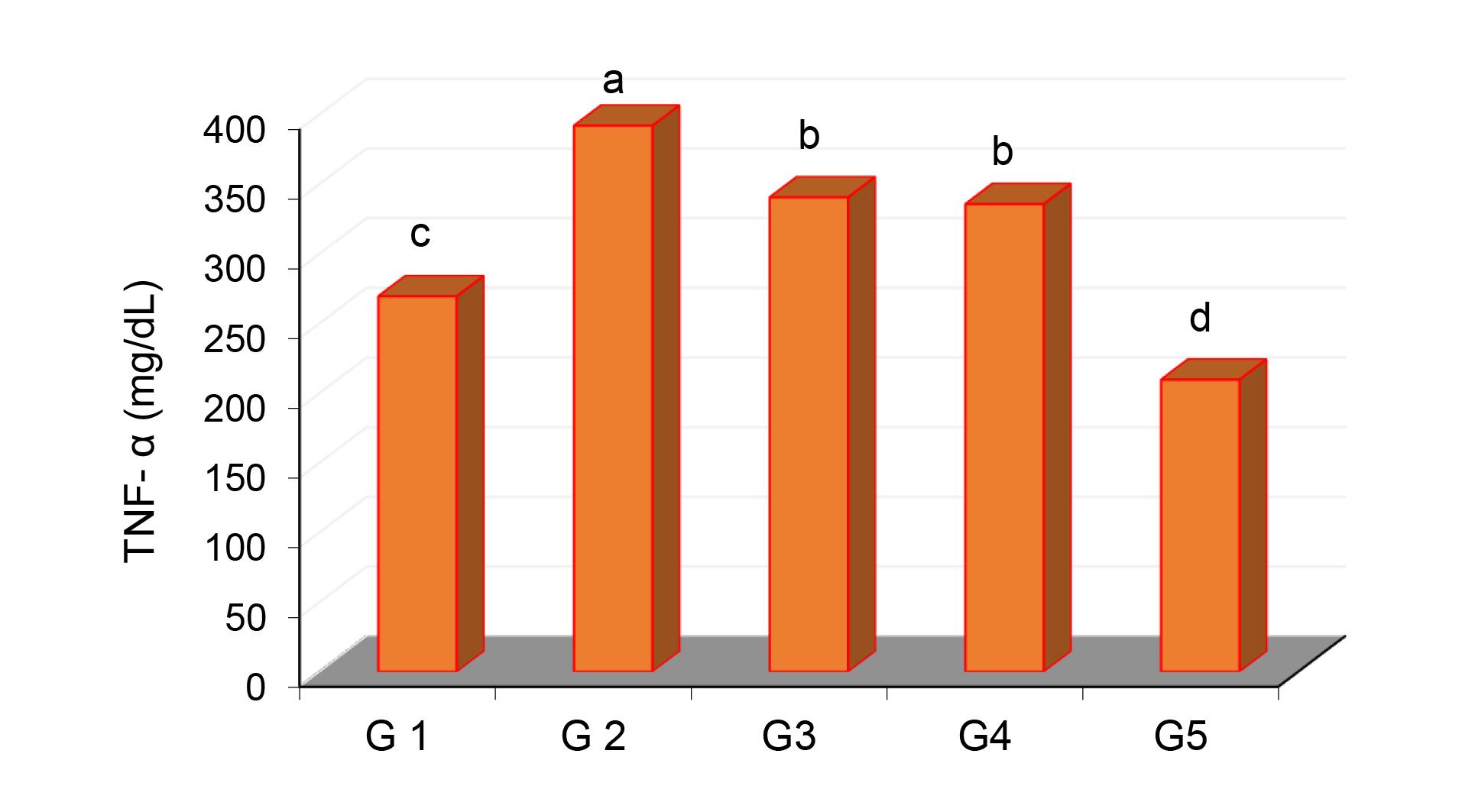
Figure 3.
Comparison between different groups regarding TNF- alpha levels. G1: control group, G2: induction group with CBZ, G3: omega-3 fatty acids with induction, G4: Vitamin E with induction, and G5: combination of omega-3 fatty acids and Vitamin E with induction. Different letters (a, b and c) indicate statistically significant differences
.
Comparison between different groups regarding TNF- alpha levels. G1: control group, G2: induction group with CBZ, G3: omega-3 fatty acids with induction, G4: Vitamin E with induction, and G5: combination of omega-3 fatty acids and Vitamin E with induction. Different letters (a, b and c) indicate statistically significant differences
Serum of IL-6 levels were also significantly elevated in group 2 (468.388 ± 22.06 U/L) compared to group 1 (268.16 ± 11.38 U/L). Regarding the treated groups (group 3, 4 and 5) showed substantial reductions in the IL-6 levels, especially in Group 5, (287.46 ± 13.57 U/L, 282.934 ± 13.95 U/L, and 160.706 ± 8.71 U/L, respectively), with P≤ 0.01. There were no significant differences between the levels of IL-6 in groups 3 and 4 (Figure 4).
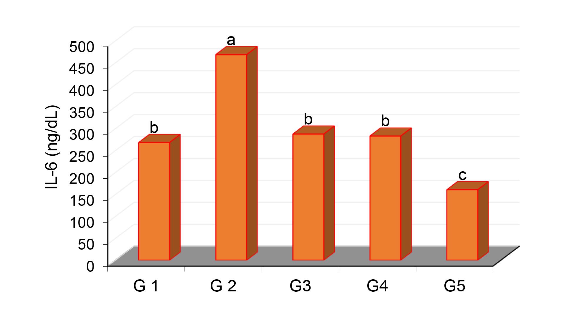
Figure 4.
Comparison between different groups regarding IL-6 levels. G1: Control group, G2: induction group with CBZ, G3: omega-3 fatty acids with induction, G4: vitamin E with induction, and G5: combination of omega-3 fatty acids and vitamin E with induction. Different letters (a, b and c) indicate statistically significant differences
.
Comparison between different groups regarding IL-6 levels. G1: Control group, G2: induction group with CBZ, G3: omega-3 fatty acids with induction, G4: vitamin E with induction, and G5: combination of omega-3 fatty acids and vitamin E with induction. Different letters (a, b and c) indicate statistically significant differences
CK-18 Gene Expression
CK-18 gene expressionwas detected in all groups as fold change, with the mean value of the control group (group 1) considered as cut off. The induction group (group 2) considered as the baseline for the comparison and exhibited the highest fold change (4.704 ± 0.51), which was approximately three times higher than the control group (1.057 ± 0.19). Groups treated with omega-3 and vitamin E (groups 3, 4 and 5) showed highly significant reductions in CK-18 expression compared to the induction group (2.511 ± 0.28, 2.782 ± 0.26, and 1.952 ± 0.17, respectively), with P < 0.01. No significant differences were observed among the treated groups (Figure 5).
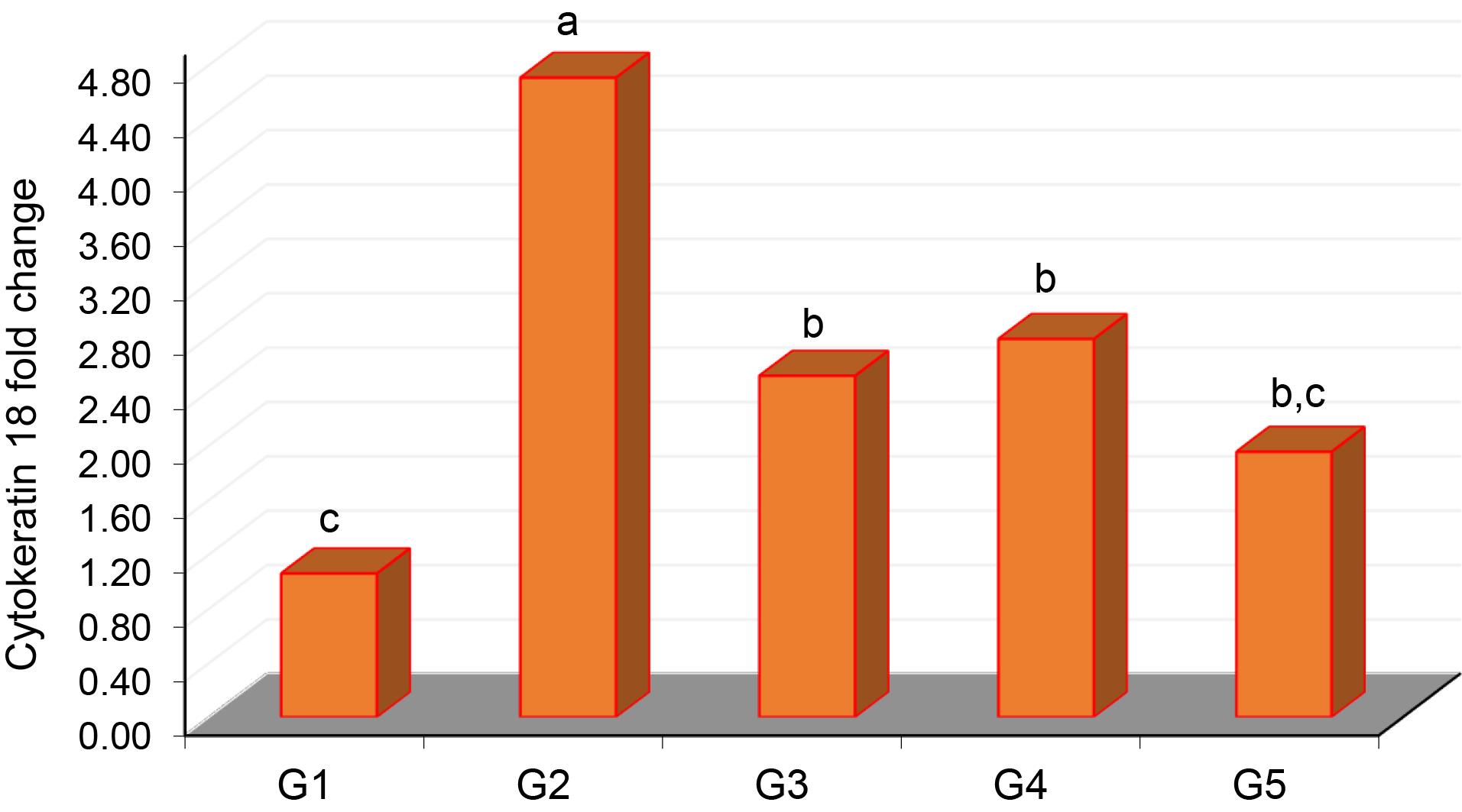
Figure 5.
Comparison between different groups regarding the mean of cytokeratin 18 gene expression (fold changes). G1: control group, G2: induction group with CBZ, G3: omega-3 fatty acids with induction, G4: vitamin E with induction, G5: combination of omega-3 fatty acids and vitamin E with induction. Different letters (a, b and c) indicate statistically significant differences
.
Comparison between different groups regarding the mean of cytokeratin 18 gene expression (fold changes). G1: control group, G2: induction group with CBZ, G3: omega-3 fatty acids with induction, G4: vitamin E with induction, G5: combination of omega-3 fatty acids and vitamin E with induction. Different letters (a, b and c) indicate statistically significant differences
Discussion
Omega-3 fatty acids and vitamin E had a potential effect on the liver; they are known for their anti-inflammatory and anti-apoptotic properties. This may offer a promising approach to protecting against liver damage during treatment with CBZ. In this study, group 2 (induced by CBZ) showed increase in all liver enzyme compared to control group, while group 3, group 4 and group 5, which included omega-3 fatty acids and vitamin E treatment, showed a significant reduction on liver enzyme levels within two weeks. In group 5, the combination of omega-3 fatty acids and vitamin E had a highly protective effect on liver cells and helped prevent hepatotoxicity.
The study results showed higher levels of ALT and ALP in group 2 which induced by CBZ, than the control group. This was in line with findings from a previous study, where ALT increased two-to three- fold and the ALP levels increased 2 to 3.5 times compared to the control group.21 The treatment groups i.e., group 3 (omega- 3 fatty acids), group 4 (vitamin E) and Group 5 (combination of omega-3 fatty acids and vitamin E) showed reductions in ALT, AST and ALP levels, bringing them closer to the control group (group 1) than to CBZ-induced group (group 2). These findings indicate that treatment alone with omega-3 or vitamin E, or in combination may provide a protective effect against CBZ-induced hepatotoxicity.
Our findings were consistent with a previous study in which treatment with omega-3 decreased ALT values in CBZ group within 1-2 weeks, approaching control group’s values.20 Recent data also indicated that omega- 3 fatty acids have a protective effect against kidney and heart toxicity.9,10
Group 2 (CBZ group) showed an increase in MDA and a decrease in GSH compared to the control group, while group 3, group 4 and group 5, which received omega-3 fatty acids and vitamin E, showed significant reductions in MDA and increases in GSH within two weeks. Especially in group 5, which combined omega-3 fatty acids and vitamin E, the effect was most highlighted. These findings aligned with those of a reported study, which noted that oxidative-stress marker MDA increased 3-fold in the CBZ group compared to the controls.20 Regarding inflammatory markers, the TNF-α level in group 2 (CBZ group) was higher than the control group. This finding is consistent with a previous study reported that CBZ treatment increased TNF-α expression.21 In our study, TNF-α levels in the treated groups (group 3, group 4) were lower than group 2 but remained slightly higher than in the control group. Meanwhile, TNF-α level in group 5 was lower than in group 2 and the other treated groups. This suggests that combination of omega-3 fatty acids and vitamin E provides stronger protection against CBZ-induced liver damage. These results align with former studies, which found that vitamin E and omega-3 fatty acids treatments reduced TNF-α expression compared to the induction group and brought levels closer to that of control.15
CK-18 expression by ELISA found that it to be a biomarker for early detection and prognosis in liver injury patients.22 In this study, CK-18 gene expression in hepatotoxicity with CBZ was also detected. Group 2 showed the highest expression, significantly greater than all other groups. In the treated groups group 3 and group 4, CK-18 expression was lower than group 2 but slightly higher than the control group. In group 5, CK-18 expression was lower than group 2 and nearly equal to the control, suggesting that omega-3 fatty acids and vitamin E -alone or combination- provided protective effects against CBZ-induced liver injury.
Conclusion
The investigation into the combination of omega-3 fatty acids and vitamin E demonstrated protective effects against CBZ-induced liver damage in rats. It revealed a multifaceted role in modulating liver enzyme levels, antioxidant status, inflammatory cytokine biomarkers, and gene expression of CK-18. These findings highlight the prophylactic potential of omega-3 fatty acids and vitamin E as a complementary therapeutic strategy to mitigate the toxic effects of CBZ treatment, promising an improvement in clinical outcomes in patients receiving this medication. Future research should explore optimal dosing, treatment duration, and potential synergistic mechanisms, as well as confirm these results through larger-scale in vivo and clinical studies.
Competing Interests
All authors declared no conflict of interest.
Data Availability Statement
All data that support the results of present study are availablein the main text.
Ethical Approval
Ethical approval was obtained from the Ethics Committee of College of Pharmacy at Mustansiriyah University for the animal study, under research number 52 and approval number 52, dated 1/10/2024.
Use of AI
The authors stated no use of AI.
References
- Baquerre C, Montillet G, Pain B. Liver organoids in domestic animals: an expected promise for metabolic studies. Vet Res 2021; 52(1):47. doi: 10.1186/s13567-021-00916-y [Crossref] [ Google Scholar]
- Allison R, Guraka A, Shawa IT, Tripathi G, Moritz W, Kermanizadeh A. Drug induced liver injury - a 2023 update. J Toxicol Environ Health B Crit Rev 2023; 26(8):442-67. doi: 10.1080/10937404.2023.2261848 [Crossref] [ Google Scholar]
- Keshari AC, Thitame SN, Aher AA, Keshari UC. Drug-induced liver injury: mechanisms, diagnosis, and management: a review. J Pharm Bioallied Sci 2025; 17(Suppl 1):S55-8. doi: 10.4103/jpbs.jpbs_568_25 [Crossref] [ Google Scholar]
- Schwarz A, Strakos C, Weihrich R. A brief review on carbamazepine: history, pharmacological properties and environmental impact. Insights in Chemistry and Biochemistry 2021; 1(4):1-4. doi: 10.33552/icbc.2021.01.000519 [Crossref] [ Google Scholar]
- Hazra D, Ellouze NF, Abri SA. Prevalence and outcomes of carbamazepine toxicity in the emergency department: a single-center retrospective study. Indian J Crit Care Med 2024; 28(9):866-70. doi: 10.5005/jp-journals-10071-24795 [Crossref] [ Google Scholar]
- Gallego MD, García MA. Acute carbamazepine intoxication. Neurol Int 2022; 14(3):614-8. doi: 10.3390/neurolint14030049 [Crossref] [ Google Scholar]
- Murty S. Antiepileptic overdose. Indian J Crit Care Med 2019; 23(Suppl 4):S290-5. doi: 10.5005/jp-journals-10071-23301 [Crossref] [ Google Scholar]
- Patted PG, Masareddy RS, Patil AS, Kanabargi RR, Bhat CT. Omega-3 fatty acids: a comprehensive scientific review of their sources, functions and health benefits. Futur J Pharm Sci 2024; 10(1):94. doi: 10.1186/s43094-024-00667-5 [Crossref] [ Google Scholar]
- Al-Hassani HK, Hummadi YA, Hatem SF. Protective effect of omega-3 and the potential of toll-like receptor gene expression in rats with doxorubicin-induced cardiac toxicity. Trop J Nat Prod Res 2023; 7(2):2358-61. doi: 10.26538/tjnpr/v7i2.9 [Crossref] [ Google Scholar]
- Al-Dabbagh AS, Mshemish BA, Al-Mugdadi SF. Investigating the potential protective effects of omega-3 against doxorubicin-induced renal injury in rats through modification of antioxidant markers. Immunopathol Persa. 2025. doi: 10.34172/ipp.2024.41707.
- Vell MS, Creasy KT, Scorletti E, Seeling KS, Hehl L, Rendel MD. Omega-3 intake is associated with liver disease protection. Front Public Health 2023; 11:1192099. doi: 10.3389/fpubh.2023.1192099 [Crossref] [ Google Scholar]
- Ara Z, Waliullah S, Rastogi D, Pant S. Vitamin E and human health: an update. Glob J Health Sci Res. 2025. doi: 10.25259/gjhsr_6_2025.
- Traber MG. Vitamin E. Adv Nutr 2021; 12(3):1047-8. doi: 10.1093/advances/nmab019 [Crossref] [ Google Scholar]
- Alorabi M, Mohammed DS, Mostafa-Hedeab G, El-Sherbeni SA, Negm WA, Mohammed AIA. Combination treatment of omega-3 fatty acids and vitamin C exhibited promising therapeutic effect against oxidative impairment of the liver in methotrexate-intoxicated mice. Biomed Res Int 2022; 2022:4122166. doi: 10.1155/2022/4122166 [Crossref] [ Google Scholar]
- Lotfi A, Mashhady Rafie S, Hesaraki S, Amini K. Protective effects of coadministration of the vitamin E and omega-3 on doxorubicin-induced hepatotoxicity in mice. Int J Cancer Manag 2022; 15(10):e121766. doi: 10.5812/ijcm-121766 [Crossref] [ Google Scholar]
- Abdou HM, Hassan MA. Protective role of omega-3 polyunsaturated fatty acid against lead acetate-induced toxicity in liver and kidney of female rats. Biomed Res Int 2014; 2014:435857. doi: 10.1155/2014/435857 [Crossref] [ Google Scholar]
- Karapehlivan M, Ogun M, Kaya I, Ozen H, Deveci HA, Karaman M. Protective effect of omega-3 fatty acid against mercury chloride intoxication in mice. J Trace Elem Med Biol 2014; 28(1):94-9. doi: 10.1016/j.jtemb.2013.08.004 [Crossref] [ Google Scholar]
- Emam H, Ahmed E, Abdel-Daim M. Antioxidant capacity of omega-3-fatty acids and vitamin E against imidacloprid-induced hepatotoxicity in Japanese quails. Environ Sci Pollut Res Int 2018; 25(12):11694-702. doi: 10.1007/s11356-018-1481-9 [Crossref] [ Google Scholar]
- Korver S, Bowen J, Pearson K, Gonzalez RJ, French N, Park K. The application of cytokeratin-18 as a biomarker for drug-induced liver injury. Arch Toxicol 2021; 95(11):3435-48. doi: 10.1007/s00204-021-03121-0 [Crossref] [ Google Scholar]
- El-Mowafy AM, Abdel-Dayem MA. Novel protection by omega-3-FAs against strychnine-induced tonic-convulsion in mice: synergy with carbamazepine. J Food Sci Nutr Res 2021; 4(3):227-39. doi: 10.26502/jfsnr.2642-11000075 [Crossref] [ Google Scholar]
- Gómez CD, Buijs RM, Sitges M. The anti-seizure drugs vinpocetine and carbamazepine, but not valproic acid, reduce inflammatory IL-1β and TNF-α expression in rat hippocampus. J Neurochem 2014; 130(6):770-9. doi: 10.1111/jnc.12784 [Crossref] [ Google Scholar]
- Cardin R, Bizzaro D, Russo FP, D’Arcangelo F, Ideo F, Pelizzaro F. Drug-induced liver injury: role of circulating liver-specific microRNAs and keratin-18. Gastroenterol Insights 2024; 15(4):1093-105. doi: 10.3390/gastroent15040075 [Crossref] [ Google Scholar]