Pharmaceutical Sciences. 31(4):440-451.
doi: 10.34172/PS.025.42799
Research Article
Preparation, Pharmaceutical Characterization, In-Vitro Release Kinetics, and Antifungal Efficacy Investigation of Fluconazole Niosomal Hydrogel
Mohammad Amin Raeisi Estabragh Data curation, Formal analysis, Investigation, Methodology, Resources, Visualization, Writing – original draft, Writing – review & editing, 1, 2, * 
Hossein Pardakhty Formal analysis, Methodology, Writing – original draft, 3
Abbas Pardakhty Conceptualization, Funding acquisition, Investigation, Project administration, Supervision, Validation, Writing – review & editing, 4, *
Author information:
1Student Research Committee, Kerman University of Medical Sciences, Kerman, Iran
2Department of Pharmaceutics, Faculty of Pharmacy, Kerman University of Medical Sciences, Kerman, Iran
3Faculty of Veterinary, Islamic Azad University, Karaj, Iran
4Pharmaceutics Research Center, Institute of Neuropharmacology, Kerman University of Medical Sciences, Kerman, Iran
Abstract
Background:
Lipid vesicular systems can enhance the penetration of antifungal drugs like fluconazole in topical applications. Niosomes, composed of non-ionic surfactants and cholesterol, are a key type of lipid vesicles.
Methods:
Fluconazole (FL) niosomes were prepared using thin film hydration with varying ratios of Span®/Tween®/cholesterol. Their morphological characteristics, particle size, physical stability, encapsulation efficiency (EE%), cumulative drug release, and kinetics were assessed. The optimal formulation was then combined with a gel base, and its physicochemical and pharmaceutical properties were examined. Antifungal efficacy against Candida albicans (ATCC: 10231) was evaluated compared to free drug solutions.
Results:
All formulations exhibited encapsulation efficiencies over 50%, with the Span60/Tween60/cholesterol blend (45/45/10 mole%) achieving the highest efficiency (70.2%). Following the Higuchi model, this formulation released 55.4% of FL over four hours. The gel formulations showed good physical stability, particularly the one with 1% carboxymethyl cellulose, which was suitable for topical application due to its pseudoplastic and thixotropic properties. In-vitro minimum inhibitory concentration (MIC) values against Candida albicans were recorded as 16 μg/mL (solution), 2 μg/mL (niosomal suspension), and 4 μg/mL (niosomal gel).
Conclusion:
A stable and locally applicable FL niosomal gel can be formulated, potentially enhancing effectiveness and reducing microbial resistance to FL as indicated by antifungal activity results in-vitro.
Keywords: Fluconazole, Niosome, Gel, Topical, Fungal, Release kinetic
Copyright and License Information
© 2025 The Author(s).
This is an open access article and applies the Creative Commons Attribution Non-Commercial License (
http://creativecommons.org/licenses/by-nc/4.0/). Non-commercial uses of the work are permitted, provided the original work is properly cited.
Funding Statement
National Institute for Medical Research and Development (Grant No.996664) supporting this research and Kerman University of Medical Sciences also support another part of the project (Grant No. 400000887).
Introduction
Fluconazole (FL) treats localized and systemic fungal infections with acceptable in vivo efficacy and pharmacokinetic properties. It has been derived from imidazole alcohol.1,2 FL inhibits ergosterol biosynthesis by inhibiting the fungal cytochrome P450 -dependent lanosterol C14α –demethylase.3 Due to FL low solubility, its bioavailability is very low.4 There are oral dosage forms commercially available, which are largely associated with some side effects, such as abdominal pain, diarrhea, flatulence, nausea and vomiting, and taste disturbance after administration.5 Topical drug administration is widely used in various medical conditions due to its numerous benefits. These include bypassing the gastrointestinal tract, avoiding gastrointestinal irritation and the hepatic first-pass effect, and directly targeting the affected area to minimize undesirable side effects.6 Unfortunately, the wide use of FL as first-line antifungal therapy has led to resistance in clinical isolates of Candida species including Candida albicans and Candida spp.2 The resistance mechanisms fungi develop to allow them to survive at higher drug concentrations. As a result, new drugs or new drug delivery systems are urgently needed to overcome this problem.7 The topical delivery of lipid vesicles provides a reliable method to administer drugs directly to the infection site, reducing drug toxicity and minimizing side effects. Enhancing drug bioavailability, particularly for poorly soluble drugs, and reducing dosage and frequency while improving patient adherence lowers the overall treatment cost.8
Niosomes, which are vesicular nanocarriers, have garnered significant attention for their potential as drug delivery systems due to their distinct advantages.9 Several routes are available for delivering niosomes, including oral, intranasal, parenteral, and topical, as well as powders, suspensions, and semisolids.10 It enhances the permeability of drugs through the skin when applied topically and enhances the oral bioavailability of poorly soluble drugs.11,12 In comparison to oily dosage forms such as ointments, and creams, aqueous suspension formulations result in better patient compliance; additionally, since niosomal dispersion is aqueous, it can be emulsified in a nonaqueous phase to regulate the drug release rate.13,14 Amphotericin B, clotrimazole, griseofulvin, ketoconazole, and itraconazole as antifungal agents are carried by niosomes.15 These developments aimed to increase drug bioavailability while minimizing adverse effects of antifungal drugs.16 The use of a mixture of surfactants (Span® and Tween®) has increased stability and percentage of entrapment, so using a mixture of two surfactants can be a more suitable option than one surfactant.17,18 The semi-solid consistency achieved through gelling agents, which are mostly cellulose derivatives and natural gums at low concentrations, reduces the formulation’s clearance rate and expands the residence period at the site of administration.19
In this research, we prepared various FL niosomal formulations, a mixture of Span® and Tween®, and evaluated them morphologically and physicochemically. Different kinetic models were assessed to determine the best in-vitro kinetic model for releasing FL from niosomes. Then, the optimal niosomes formulation incorporated in the gel base and its physicochemical properties were evaluated. Finally, in-vitro, the antifungal effect of selected niosomal gel formulation against Candida albicans (ATCC:10231) was studied.
Materials and Methods
Materials
FL was a kind gift from Dr Kiafar (Zahravi Pharmaceutical Co., Tabriz, Iran). Span® 20, 40, and 60 and Tween® 20, 40, and 60 as surfactants from Fluka Company (Switzerland), and cholesterol was obtained from Sigma-Aldrich Company (USA). Culture medium, polymers, all organic solvents, and chemicals were purchased from Merck Chemical Company (Germany).
Preparation of FL niosomes
The thin-layer film hydration method was used to prepare FL niosomes.20 Various niosomal formulations containing FL, surfactants, and cholesterol were prepared (The total concentration of the lipid phase in all formulations was 60 μM presented in Table S1 (Supplementary file). As the organic solvent, chloroform was used to dissolve FL (final concentration: 5 mg/mL), surfactants, and cholesterol. Rotating evaporators (Heidolph, Germany) were used to remove chloroform. After the lipid film was dried, distilled water was added and rotated for 30 min at 180 rpm and 60 °C. For complete hydration, the formed lipid vesicles were stored at room temperature for 24 h in borosilicate glass vials and then in a refrigerator (2-8 °C) for future studies. Also, the separation of the drug entrapped in the niosomes and the free drug was performed using the dialysis method.
Morphologic study, size and zeta potential analysis of niosomal formulations
The niosomes were examined for shape, aggregation of vesicles, and separation of constituents as morphological characteristics by a light microscope (Leitz, HM-LUX3, Germany) equipped with a digital camera with × 400 magnification. Mean volume diameters, and vesicular size distribution of vesicles were calculated by static laser light scattering (Malvern MasterSizer 2000E, UK) technique.21 Also, the zeta potential of the formulation was measured one week after its manufacture (Malvern Zetasizer Pro, UK). The range of dispersity (span), which can vary from almost monodisperse to highly polydisperse, was determined using the following equation:
In which dV90, dV50, and dV10 are cumulative 90, 50, and 10 percent undersize volume size distributions, respectively.22
Determination of FL concentration
The quantity of FL was measured using UV spectrophotometry.23 The FL standard solution (600 µg/mL) was created in an 80:20 v/v ethanol and water mixture, and it was scanned using a UV spectrophotometer (UV/Visible Spectrophotometer Optizen 3220, South Korea) at a wavelength of 200 to 400 nm. Standard FL solutions (50-600 µg/mL) were created once the maximum absorption wavelength (λmax) was established, and the absorption of these solutions was measured at the λmax. Using Microsoft Office Excel® software, the absorption versus concentration graph was created, and its equation was derived.
Encapsulation efficiency percent (EE%)
To calculate the EE% of FL the dialysis method was used.24 Unencapsulated drugs were removed from the niosome suspensions using the dialysis method. To do this, one milliliter of the niosome suspension was placed into a dialysis bag (Visking tube, with a molecular weight cut off of 12 KD) and dialyzed against a mixture of ethanol and water (80:20 v/v%) for four hours.22,25 This process effectively removed any unbound drug, leaving only the encapsulated drug within the niosome. Drug concentration was determined spectrophotometrically at 260 nm. The percentage of the drug encapsulated was calculated using the following equation:
where Cencap and CF denote the amount of FL encapsulated in niosomes and the free amount of FL, respectively.
In vitro release kinetic of FL from niosomes
Franz diffusion cell (Ashk-Shishe, Iran, Receptor volume:15 mL) and cellulose acetate dialysis tube (Visking tube, MW cut off 12 KD) as the membrane was used to study the release.26 The receptor phase is a mixture of ethanol and water (80:20 v/v%) and sampling is done at different times. After determining the concentration, the graph of the cumulative released percentage of FL is drawn against time. To determine the release kinetics of FL for the optimal formulation, the in-vitro drug release data were fitted using a data-fitting solver in Excel® software (Microsoft Office, 2016) with different release kinetic models and evaluated to understand the drug release kinetics. These included a zero-order, a first-order, Higuchi, the Korsmeyer-Peppas, and Hixon Crowell’s models.27
Preparation of niosomal gels
In this research, Carbomer 940®, carboxymethyl cellulose (CMC), and hydroxypropyl methylcellulose (HPMC) were used as gelling agents in different percentages (Table 1). In formulations containing Carbomer 940®, 3-5 drops of triethanolamine will be added to prepare a clear gel. Different amounts of polymer were dispersed in water and with the help of a stirrer with a constant speed of 2000 rpm, stirring continued for 30 minutes until uniformity at 25 ºC, then the niosome suspension was added and mixed well. The obtained FL niosomal gel was 0.25%.
Table 1.
The percentage of components of FL niosomal gel formulations
|
Formulation name
|
Gelling agent
|
Gelling agent (%)
|
Propylene glycol (%)
|
| FNG1 |
CMC |
1 |
- |
| FNG2 |
2 |
- |
| FNG3 |
Carbomer 940 |
1 |
10 |
| FNG4 |
2 |
10 |
| FNG5 |
HPMC |
2 |
- |
| FNG6 |
4 |
- |
Physicochemical evaluation of niosomal Gels
Physical appearance
The prepared formulations were checked visually regarding appearance characteristics including transparency, color, absence of air bubbles, and uniformity.28
Physicochemical stability
To check the physical stability in various temperatures, 15 g of each formulation was placed at -8°C for 48 h and then at 25°C for 48 h. This process was repeated for 6 cycles and the results were evaluated and reported based on the amount of leakiness, wrinkling, liquid seepage, color change, and bubble creation.29 Also, to evaluate the chemical stability of FL, the selected formulation was stored at four temperatures (-8, 4,25, and 40 °C) and the amount of FL was determined by UV spectrophotometry at one week, one month, three, and six months.30
pH of formulation
One g of the prepared formulation was dispersed in 15 ml of water. pH was measured three times and reported as an average. This measurement was done at zero, 48 h, 1 week, 1 month, and 6 months after preparation of formulation (stored at room temperature).31
Drug content
To check the content uniformity of the gel formulation, one g of the formulation was weighed and dispersed in 5 ml of water, and then 20 ml of ethanol was added to it and the amount of FL was calculated.31
Rheological behaviors
The rheological behaviors test was done with a Brookfield rheometer (Brookfield Engineering, USA). The gel formulation was poured into the beaker and using the LV4 spindle at room temperature, the spindle started rotating at different speeds (at first increasing and then decreasing) and the opposite torque force was measured at each speed. Then, a graph of revolutions per minute was drawn against the opposite torque force.32
In-vitro FL niosomal gel release
The release rate of FL from niosomal gel was performed using a Franz diffusion cell and the same method as described in section 2.5.
In-vitro antifungal effects study
Antifungal activity in the laboratory environment of FL solution (as a control), empty niosome gel, FL niosome suspension, and FL niosome gel against Candida Albicans (ATCC:10231) was examined and the minimum inhibitory concentration (MIC) was calculated. Different amounts of the above formulations were added to tubes containing one ml of Sabouraud Dextrose Broth culture medium, and then concentrations (0 to 128 μg/mL) of FL were prepared by serial dilution method. Then, 0.2 ml of microbial suspension with turbidity equivalent to 1.5 × 10⁸ CFU/mL was added to tubes. Also, two tubes were considered as positive and negative control. After 4 days of incubation at 37 °C, the tubes were checked visually for turbidity.33
Statistical analysis
Statistical analysis was carried out using IBM SPSS Software (Version 26, IBM, USA). Group comparisons were performed through a one-way ANOVA, followed by Tukey’s Multiple Comparison test, with the significance threshold set at P <0.05.
Results
Determination of FL
Using the UV spectrophotometer, the λmax of FL (in ethanol) was equal to 260 nm. This finding was consistent with the previous studies.34 The equation of the line is:
Absorbance = 0.0019 Concentration – 0.0118 R2 = 0.9989
Morphology and size analysis
After preparing different niosomes formulations, the shape of niosomes and the presence of crystals in them were checked using an optical microscope. All formulations produced niosomes, however some formulations such as F4 and F7 showed a significant amounts of likely crystal. Figure 1 shows the light microscope photo of F7 and F9.
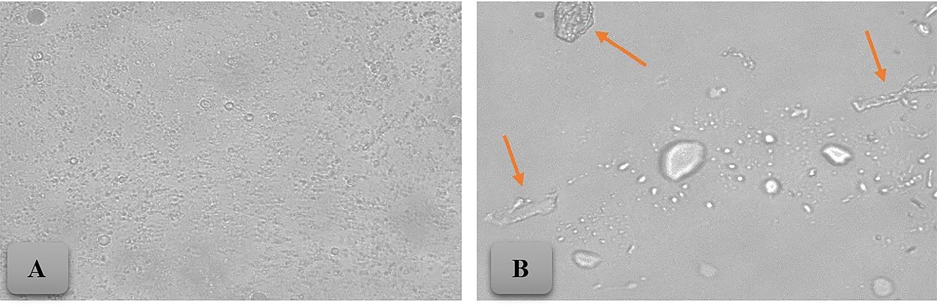
Figure 1.
Optical micrographs of FL niosomes (× 400 magnification): (A) F9, (B) F7 (The arrow indicates the presence of likely crystals)
.
Optical micrographs of FL niosomes (× 400 magnification): (A) F9, (B) F7 (The arrow indicates the presence of likely crystals)
Figure 2 shows the size distribution diagram of niosomal formulations. Based on the morphology of niosomes, the absence of crystals, and the vesicle size distribution diagram, formulations F2, F3, F6, and F9 were selected for further study. The range of dispersity (span) of thesis formulation is 3.48, 1.85, 2.73, and 1.31, respectively. Also, Table 2 shows the volumetric diameter (dv), span, zeta potential and EE percent of the prepared formulations.
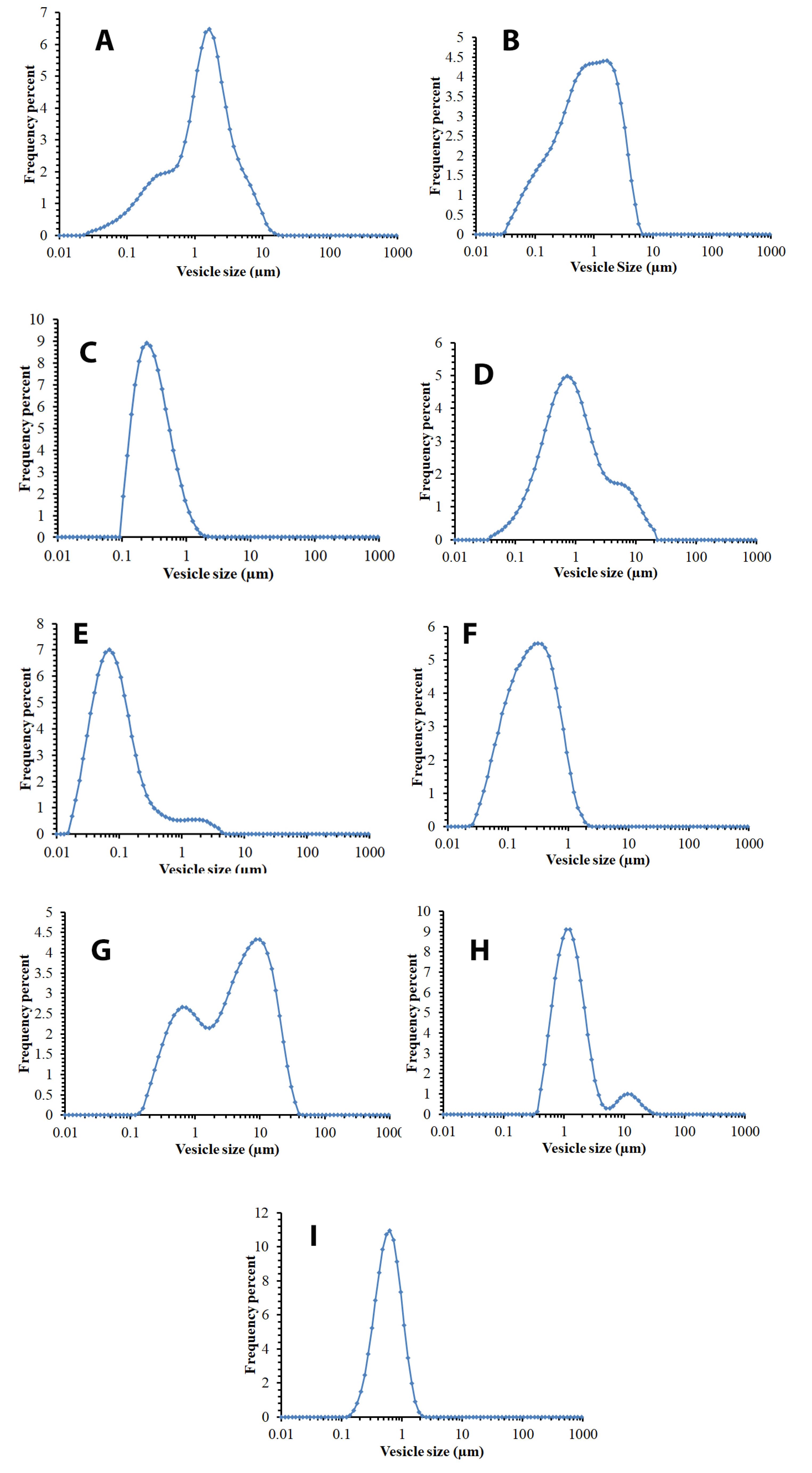
Figure 2.
The vesicle size distribution diagram of niosome formulations one week after preparing and storage at 2-8ºC. A: F1 (Span20/Tween20/cholesterol 25/25/50 mole%), B: F2 (Span20/Tween20/cholesterol 35/35/30 mole%), C: F3 (Span20/Tween20/cholesterol 45/45/10 mole%), D: F4 (Span40/Tween40/cholesterol 25/25/50 mole%), E: F5 (Span40/Tween40/cholesterol 35/35/30 mole%). F: F6 (Span40/Tween40/cholesterol 45/45/10 mole%), G: F7 (Span60/Tween60/cholesterol 25/25/50 mole%), H: F8 (Span60/Tween60/cholesterol 35/35/30 mole%), I: F9 (Span60/Tween60/cholesterol 45/45/10 mole%)
.
The vesicle size distribution diagram of niosome formulations one week after preparing and storage at 2-8ºC. A: F1 (Span20/Tween20/cholesterol 25/25/50 mole%), B: F2 (Span20/Tween20/cholesterol 35/35/30 mole%), C: F3 (Span20/Tween20/cholesterol 45/45/10 mole%), D: F4 (Span40/Tween40/cholesterol 25/25/50 mole%), E: F5 (Span40/Tween40/cholesterol 35/35/30 mole%). F: F6 (Span40/Tween40/cholesterol 45/45/10 mole%), G: F7 (Span60/Tween60/cholesterol 25/25/50 mole%), H: F8 (Span60/Tween60/cholesterol 35/35/30 mole%), I: F9 (Span60/Tween60/cholesterol 45/45/10 mole%)
Table 2.
Composition, and size of different niosomal formulations containing FL (5 mg/mL)
|
Name
|
Constituents of the lipid phase
|
Molar ratio
|
Volumetric diameter (µm)
|
span
|
Zeta potential (mv)
|
EE%
|
|
dv10%
|
dv50%
|
dv90%
|
| F 1 |
Span20/Tween20/Cholesterol |
25/25/50 |
0.623 |
4.073 |
13.111 |
3.07 |
-15.11 |
ND |
| F 2 |
Span20/Tween20/Cholesterol |
35/35/30 |
0.384 |
2.339 |
8.527 |
3.48 |
-18.22 |
63.7 |
| F 3 |
Span20/Tween20/Cholesterol |
45/45/10 |
0.154 |
0.303 |
0.717 |
1.85 |
-22.17 |
53.9 |
| F 4 |
Span40/Tween40/Cholesterol |
25/25/50 |
0.762 |
3.067 |
20.657 |
6.48 |
-19.33 |
ND |
| F 5 |
Span40/Tween40/Cholesterol |
35/35/30 |
0.741 |
1.836 |
8.215 |
1.07 |
-21.22 |
ND |
| F 6 |
Span40/Tween40/Cholesterol |
45/45/10 |
0.178 |
0.615 |
1.858 |
2.73 |
-29.88 |
66.3 |
| F 7 |
Span60/Tween60/Cholesterol |
25/25/50 |
1.122 |
10.081 |
40.103 |
3.87 |
-48.33 |
ND |
| F 8 |
Span60/Tween60/Cholesterol |
35/35/30 |
1.778 |
3.566 |
10.982 |
2.59 |
-45.11 |
ND |
| F 9 |
Span60/Tween60/Cholesterol |
45/45/10 |
1.435 |
2.770 |
5.062 |
1.31 |
-36.22 |
70.2 |
Encapsulation efficiency percent
In this study, the EE% of the prepared niosomal formulations was above 50%, and the highest EE% was related to F 9 with a rate of 70.2. The EE% of the selected formulations is shown in Table 2.
In vitro release from niosomes
Figure 3 displays the FL release of the selected formulations. The data of the study of the release kinetics of the formulations can be found in Table 3. The highest release in niosomal formulations after 4 h is related to F6 with a value of 57.6%. Also, the release percentage in F9 is 55.4 and the release kinetic follows the Higuchi model (R2 = 0.975, k = 3.625).
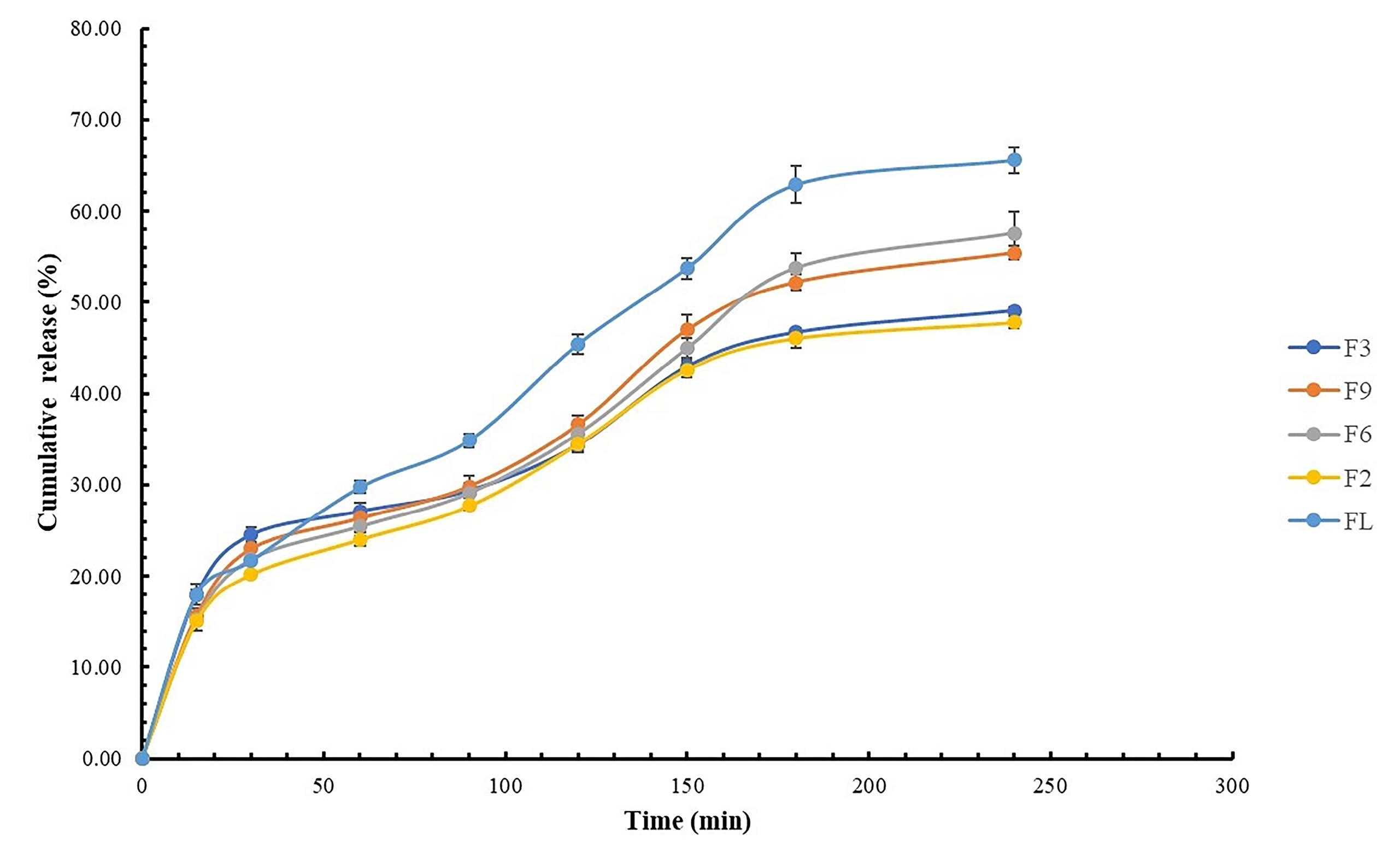
Figure 3.
The release profile of FL solution (FL), and selected FL niosomal suspension (mean ± SD, n = 3, P value = 0.0367)
.
The release profile of FL solution (FL), and selected FL niosomal suspension (mean ± SD, n = 3, P value = 0.0367)
Table 3.
Result of in vitro kinetic release study profile of FL niosomes and niosomal gel
|
Formulation
|
Zero-order
|
First order
|
Higuchi
|
Peppas
|
Hixon-Crowell
|
|
K
|
R2
|
K
|
R2
|
K
|
R2
|
K
|
R2
|
n
|
K
|
R2
|
| FL |
0.330 |
0.933 |
0.005 |
0.970 |
4.254 |
0.978 |
0.041 |
0.968 |
0.503 |
0.007 |
0.963 |
| F2 |
0.251 |
0.893 |
0.003 |
0.934 |
3.251 |
0.979 |
0.044 |
0.966 |
0.435 |
0.005 |
0.923 |
| F3 |
0.258 |
0.853 |
0.003 |
0.911 |
3.367 |
0.965 |
0.067 |
0.945 |
0.358 |
0.004 |
0.894 |
| F6 |
0.283 |
0.933 |
0.004 |
0.963 |
3.622 |
0.970 |
0.040 |
0.952 |
0.476 |
0.005 |
0.957 |
| F9 |
0.281 |
0.912 |
0.004 |
0.953 |
3.625 |
0.975 |
0.045 |
0.957 |
0.445 |
0.005 |
0.942 |
| FNG1* |
0.190 |
0.892 |
0.002 |
0.925 |
2.545 |
0.990 |
0.023 |
0.984 |
0.514 |
0.003 |
0.915 |
* FNG1 is the gel formulation (Table 1) of selected FL niosome (F9).
Physicochemical properties of niosomal Gels
Physical appearance
Due to the appropriate morphology, zeta potential, normal vesicle size distribution, and high EE%, the F9 formulation was chosen for further study and preparation of the niosomal gel. Formulations containing CMC had the most uniformity (FNG1, and FNG2). On the other hand, the formulation containing Carbomer 940® had the most air bubbles (FNG3, and FNG4). The gels containing Carbomer were transparent and light yellow (FNG3, and FNG4), while CMC and HPMC gels were transparent and colorless, tending to white (FNG1, FNG2, FNG5, and FNG6). Regarding adhesion, the gel containing 2% Carbomer (FNG3) had the highest apparent stickiness.
Physicochemical stability
To assess the shelf life of the niosomal gel formulations, they were examined after undergoing 6 thermal cycles. Most formulations were found to be stable, with no noticeable physical or organoleptic changes. However, the formulations containing Carbomer were associated with increased air bubbles, and some shrinkage was also observed in them. The formulation of selected niosomal gel (FNG1) was stable at three temperatures (-8, 4, and 25 ºC) for at least six months, but at a temperature of 40 degrees after 3 months, the percentage of the remaining drug was less than 90% and instability was observed. The graph in Figure 4 displays the percentage of remaining FL at various temperatures and time intervals (Table S3), serving as a means to evaluate the shelf life of FNG1.
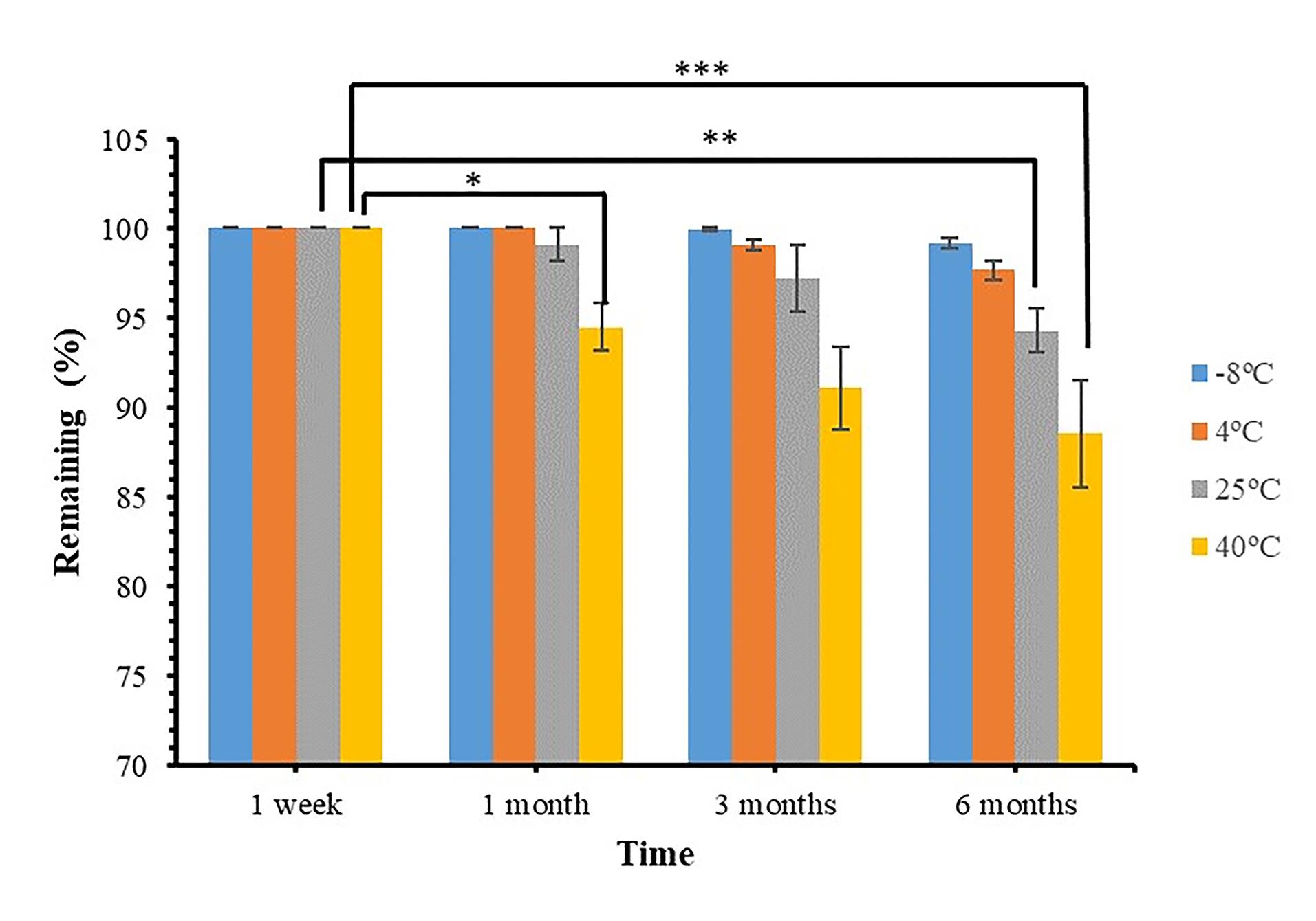
Figure 4.
The remaining drug percentage at different temperatures and times for FNG1 formulation (Mean ± SD, n = 3, * (P < 0.05), ** (P < 0.001), *** (P < 0.0001)).
.
The remaining drug percentage at different temperatures and times for FNG1 formulation (Mean ± SD, n = 3, * (P < 0.05), ** (P < 0.001), *** (P < 0.0001)).
pH of formulation
The formulation containing Carbomer (FNG3, and FNG4) had a higher pH than other formulations (7.71, and 7.02), which could be due to the use of triethanolamine to increase the pH of the environment and form a gel.35,36 The pH level of all formulas was suitable for topical use (5.80-7.71) and over time, small changes were observed in the form of a decrease in pH, which could be due to the absorption of CO2 from the air and its reaction with water in the formulation.37 The results of the pH measurement and its changes over six months are presented in Supplementary file (Table S2).
Drug content
The content uniformity test results are presented in a Supplementary file (Table S2). Based on the results, all the formulations had uniformity above 90%. Formulations containing Carbomer 940® exhibited the lowest content uniformity, potentially attributed to the presence of air bubbles and its rapid gelation upon adding triethanolamine.
Rheological behavers
The gel formulation containing Carbomer 940® contained several air bubbles after adding triethanolamine and gelling. Also its drug content was less uniform and was excluded from the study. The rheological behavior of niosomal gels containing CMC and HPMC was investigated. The CMC gel showed more pseudoplastic and thixotropic behavior than the HPMC gel, making it a more suitable candidate for topical use.38,39 Also, 2% CMC gel had high viscosity, and its 1% formula (FNG 1) with appropriate viscosity was selected for further study.40,41 Figure 5 shows the results of investigating the rheological behavior of the prepared formulations.
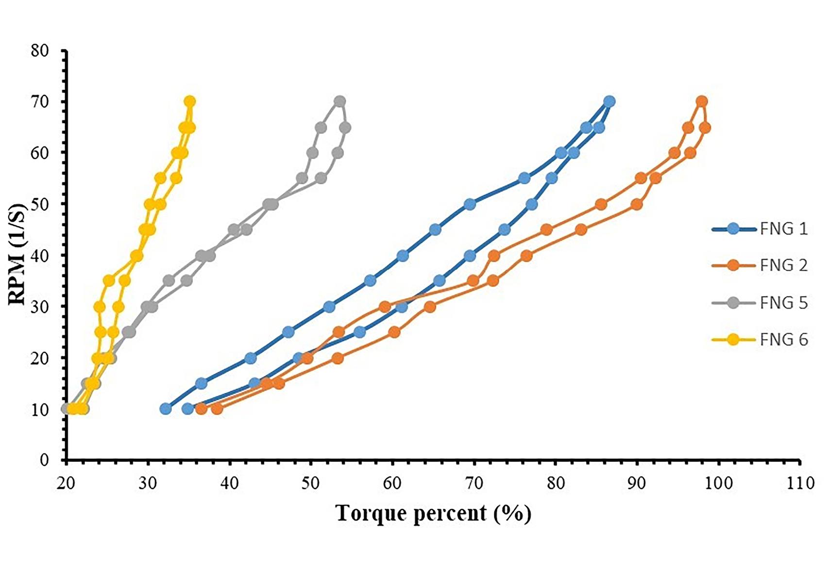
Figure 5.
The results of investigating the rheological behavior of the selected niosomal gel formulations.
.
The results of investigating the rheological behavior of the selected niosomal gel formulations.
In vitro FL niosomal gel release
The release profile of FL niosomal gel (FNG1) is presented in Figure 6. After 4 h, the in vitro release of this formulation was 35.9%. Although the release rate constant of FL from niosomal gel is lower than that of niosomal suspension, it fits the Higuchi model.
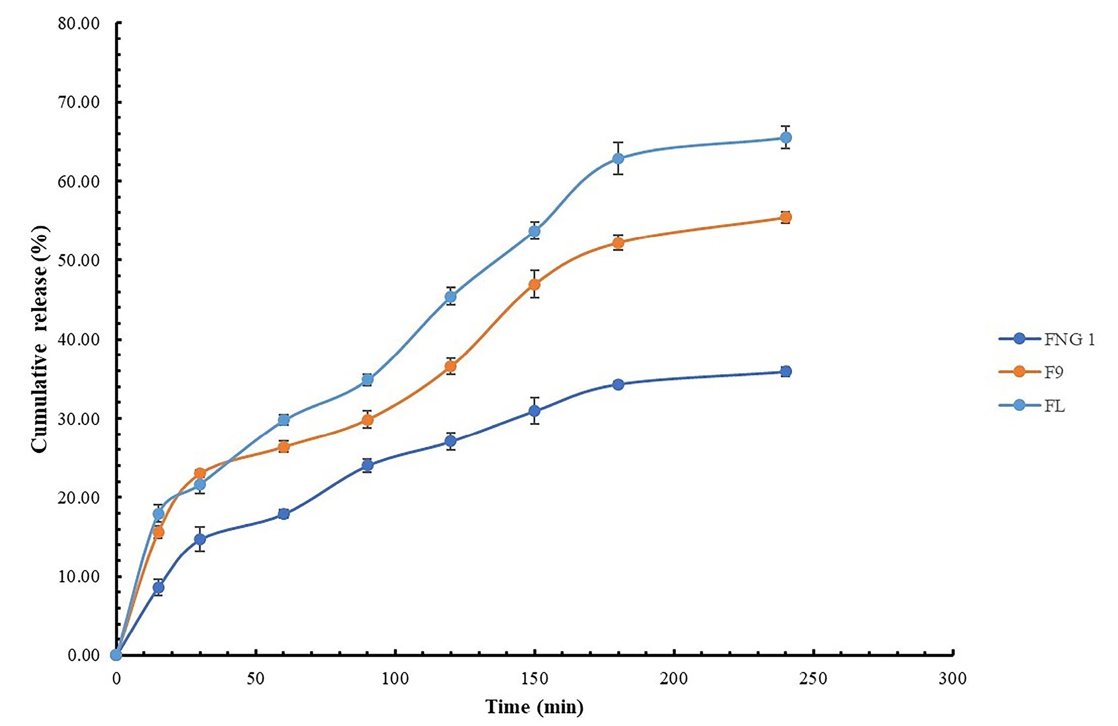
Figure 6.
The release profile of FL solution (FL), FL niosomal suspension (F9), and FL niosomal gel (FNG1) (Mean ± SD, n = 3, P = 0.0231)
.
The release profile of FL solution (FL), FL niosomal suspension (F9), and FL niosomal gel (FNG1) (Mean ± SD, n = 3, P = 0.0231)
In-vitro antifungal effect
Based on previous physicochemical tests, the niosomal formulation (F9) and the niosomal gel formulation (FNG1) were selected to investigate the antifungal effect. A drug-free niosomal gel formulation with similar compositions to FNG1 was prepared and studied as a control. The MIC of the prepared formulations against Candida albicans (ATCC: 10231) is reported in Table 4. The use of lipid bilayer drug delivery systems, such as liposome and niosome, leads to increased antifungal effects and is one of the solutions to overcome microbial resistance. In past studies, including the Musavi et al study, the use of FL liposome has increased its antifungal effects.42 Also, in other studies, using niosome increased the antifungal effects.15,43 The use of gel-based formulations, considering that it is more acceptable to the patient and increase the stability, can improve the effectiveness of the topical medicinal product.44 In this study, investigating the antifungal effects of FL niosomal formulations in the laboratory environment showed a decrease in the MIC, which is consistent with previous studies.45,46 Niosomal gel formulation had a higher MIC than niosomal suspension, which could be due to the role of the gel base in reducing the release rate of FL.47
Table 4.
The MIC of the prepared formulations against Candida albicans (ATCC: 10231)
|
Formulation name
|
Content of the formulation
|
MIC (µg/mL)
|
| FL |
FL solution |
16 |
| ENG |
Niosomal gel without drugs |
- |
| F9 |
FL niosomal suspension |
2 |
| FNG1 |
FL niosomal gel |
4 |
Discussion
Niosomes were formed in all formulations, large amount of likely crystal was observed in some formulations, including F4 and F7. This issue can be due to the competition between cholesterol and FL as the lipophilic drug to be placed in the lipid bilayer. Past studies have used a mixture of multiple surfactants to achieve the required HLB.26,48,49 Cholesterol and ergosterol are also used to increase the stability of the lipid bilayer in specific proportions.50,51 More niosomes were observed in the formulations containing Span20/Tween20/cholesterol and Span60/Tween60/cholesterol than in the formulation containing Span40/Tween40/cholesterol. In the thin layer hydration method, niosomes are mainly formed MLV.52 In this study, F5, F6, and F9 were mainly MLV.
Vesicle size distribution is one of the important physicochemical properties in the preparation and optimization of niosomes, which plays a role in them in vitro and in vivo effects. Cholesterol one of the components used for different reasons such as double layer stabilizer, phase change temperature regulator, and drug release controller, can affect the vesicle size.53 In the present study, it was observed that as the molar percentage of cholesterol increased, there was a corresponding increase in the size of niosomes. It has also been observed in the preparation of insulin niosomes.54,55 Due to the lipophilic structure of FL and structural similarity with ergosterol and cholesterol, the presence of FL can also lead to changes in vesicle size in niosomes. In the study of Shirsand et al, the presence of ketoconazole in the structure of niosomes led to a change in the vesicle size of niosomes.47 Figure 3 illustrates that as cholesterol concentrations increase (from F9 to F7 and F6 to F4), there is a corresponding increase in the frequency of larger vesicles. This can be attributed to the higher levels of cholesterol, which leads to larger area per molecule and a thinner bilayer.56 The observed pattern, with two distinct peaks at approximately 1 µm and 10 µm, which may indicate the aggregation of niosomes (F4 and F7). Formulation F9 has a bell-shaped and appropriate vesicle size distribution, while the formulations with a high percentage of cholesterol have an asymmetric vesicle size distribution. In Span20/Tween20/Cholesterol and Span40/Tween40/Cholesterol formulations, increasing cholesterol decreases negative charge density and zeta potential, as noted in prior studies.57,58
FL has a lipophilic structure similar to ergosterol and cholesterol. Niosome, as a lipid bilayer structure, can potentially trap it in its bilayer. The Mousavi et al study, a similar EE% was observed for FL liposome.42 The results of this study show a higher EE% for FL compared to the study of Gupta et al, which can be due to our use of a mixture of two surfactants (Span® and Tween®), which leads to the formation of suitable and more stable structures.59 In the Span20/Tween20 formulation, the EE% and particle size increased as the cholesterol percentage increased. However, due to the strong hydrophilicity (high HLB) of the Span20/Tween20 formulation, prevented a significant change in the bilayer’s hydrophobicity despite the increase in cholesterol percentage. As a result, there were no significant changes in EE%. This could be due to the larger size of the niosomes, which allows for a greater amount of FL to be incorporated into the lipid bilayers.60,61 In general, with the increase in surfactants’ hydrocarbon chain length the EE% of water-insoluble drugs such as alpha-lipoic acid25, carvedilol,62 and flurbiprofen63 increases. This study also observed this result for the surfactants Span20/Tween20, Span40/Tween40 and Span60/Tween60. As the HLB increases, the entrapment percentage of lipophilic drugs decreases. This phenomenon has also been observed in the study of curcumin niosomes. This could be attributed to the lower affinity of surfactants with higher HLB values for lipophilic molecules.58 The EE% of the selected formulations is shown in Figure 4. The use of nonionic surfactants and encapsulation of the drug in the niosome structure leads to increased solubility of poorly soluble drugs.60 Aldosari et al utilized various concentrations of different nonionic surfactant as stabilizers to create FL nanosuspension formulations. The impact of these stabilizers on the particle size and zeta potential of the nanosuspensions varied depending on the type and concentration of the stabilizer used. The solubility of the nanosuspension formulations improved by up to 5.7 times compared to the untreated drug.64
In the study by El-Ridy et al, the mechanism of drug release was found to follow the Higuchi model in the niosome formulation.65 Higuchi created various theoretical models to analyze the release of drugs soluble in water or have low solubility in semi-solid or solid forms. Mathematical equations were derived for drugs dispersed in a consistent matrix that behaves like the diffusion medium. Higuchi explains that drug release is a diffusion process dependent on the square root of time and based on Fick’s law principle.27 This correlation can be applied to describe the dissolution of drugs from various types of modified-release pharmaceutical dosage forms, including transdermal systems. It is important to ensure the validity of this relationship both during the initial release of the nonencapsulated drug and before the drug delivery carrier degrades and affects the diffusion coefficient. The Higuchi equation can be used to determine the apparent diffusion coefficient of a drug in a drug delivery system. This equation only has one unknown diffusion coefficient during the release period. This coefficient can be calculated through nonlinear regression using experimental data for controlled release formulations.66 In cases such as the release of alpha lipoic acid,25 caffeine,67 insulin,68 and carvedilol,62 the drug release curve is biphasic. In this project, a biphasic release diagram was observed for niosome formulations, which could be because at first, small amounts of the drug are free or in the form of surface absorption of niosomes; It causes a release with a higher gradient, and then the FL trapped in the niosome structure is gradually released with a lower gradient from the membrane. Also, based on the Korsmeyer and Peppas models and n value the release of the FL from niosomal formulation (F9) follows Fick’s law ( ≤ 0.45) and diffusion is the main release mechanism.27 In the study conducted by El-Housiny et al, FL solid lipid nanoparticles were prepared using varying concentrations of solid lipids (Compritol 888 ATO, Precirol ATO5) and surfactants (Cremophor RH40, Poloxamer 407). The Higuchi equation best describes the drug release pattern from almost all SLN formulations, which explains the diffusion of the drug from homogeneous and granular matrix systems.69
The release of FL from niosomal gel also follows the Higuchi model, but its release rate constant has decreased compared to niosomal suspension. This shows the role of the gel in reducing the initial release of unentrapped drugs in the niosome. In the study of Moghassemi et al, this issue was also observed in the release from niosomal gel.54 Also in Akbarzadeh et al study on the release of simvastatin70 and Garg et al study on the release of the antifungal drug luliconazole,60 niosomal gel formulation followed the Higuchi model.
Encapsulation of terbinafine as an antifungal agent in niosomal structure increased its effectiveness against Aspergillus, Fusarium, and Trichophyton.71 Niosomal formulation of antifungal drugs can be delivered transdermally instead of orally.72,73 Recently, Yasin et al studied, contact lenses containing FL niosomes that were prepared using Span 60 and cholesterol. The statistical analysis revealed that contact lenses containing FL-loaded niosomes exhibited a significantly greater reduction in fungal adhesion than contact lenses containing only FL.74 In the study conducted by Fatima et al, nano vesicular carriers were created using the ether injection method. These carriers were composed of varying levels of cholesterol, combined with free fatty acids and a monoester of polyoxyethylene fatty. The aim of the study was to investigate the antifungal effect of FL vesicles on C. albicans. The results showed improved efficacy and reduced MIC values compared to the FL solution.75 In past studies, including the Musavi et al study, the use of FL liposome has increased its antifungal effects.42 In the study conducted by Agarwal et al, a FL niosomal gel was prepared using only Span 60 or Tween 60. The resulting niosomes were found to be spherical and their release percentage was examined. However, the study did not investigate the gel’s rheological properties or the final formulation’s antifungal effectiveness.4 The use of gel-based formulations, considering that it is more acceptable to the patient and also increase the stability, can lead to the improvement of the effectiveness of the topical medicinal product.44 In this study, investigating the antifungal effects of FL niosomal formulations in the laboratory environment showed a decrease in the MIC, which is consistent with previous studies.45,46 Niosomal gel formulation had a higher MIC than niosomal suspension, which could be due to the role of the gel base in reducing the release rate of FL.47
Conclusion
Based on the physicochemical evaluation of the prepared formulations, it is possible to create a stable and effective niosomal FL gel by using a combination of Span 60/Tween 60/cholesterol (45/45/10 mole%) as the components of the niosome and 1% CMC as a gelling agent. In laboratory testing, this formulation showed a MIC of 4 µg/mL, while the FL solution had a MIC of 16 µg/mL. This suggests that the niosomal gel could be used as a drug delivery system to enhance the effectiveness of FL. One limitation of this study is that it only examined the antifungal effect on a single strain of Candida and did not include any animal studies. Further studies should be conducted to determine the impact of the niosomal gel on various fungal species, such as dermatophytes. If the results are promising, this formulation could be considered for clinical trials as a topical treatment for fungal diseases in both animals and humans.
Competing Interests
The authors declare that have no competing financial interests or personal relationships that could have appeared to influence the work reported.
Ethical Approval
In this study, the principles of ethics in research have been fully observed and approved by the Research Ethics Committees of National Institute for Medical Research Development (IR.NIMAD.REC.1399.227) and Research Ethics Committee of Kerman University of Medical Sciences (IR.KMU.REC.1401.061).
Supplementary Files
Supplementary file 1 contains Tables S1-S3.
(pdf)
Acknowledgements
A part of this article is the result of a research project, which was carried out with the financial support of the National Institute for Medical Research and Development (Grant No.996664). Also, we thank the Kerman University of Medical Sciences for supporting another part of the project (Grant No. 400000887).
References
- Syed SM, Gaikwad SS, Wagh S. Formulation and evaluation of gel containing fluconazole microsponges. Asian J Pharm Res Dev 2020; 8(4):231-9. doi: 10.22270/ajprd.v8i4.753 [Crossref] [ Google Scholar]
- Emami S, Ghobadi E, Saednia S, Hashemi SM. Current advances of triazole alcohols derived from fluconazole: design, in vitro and in silico studies. Eur J Med Chem 2019; 170:173-94. doi: 10.1016/j.ejmech.2019.03.020 [Crossref] [ Google Scholar]
- Bongomin F, Oladele RO, Gago S, Moore CB, Richardson MD. A systematic review of fluconazole resistance in clinical isolates of Cryptococcus species. Mycoses 2018; 61(5):290-7. doi: 10.1111/myc.12747 [Crossref] [ Google Scholar]
- Agarwal S, Misra R, Poddar RK, Yadav VK. Formulation and in vitro evaluation of fluconazole niosomal gel for topical drug delivery. J Pharm Negat Results 2023; 13(8):3963-75. doi: 10.47750/Pnr.2022.13.S08.498 [Crossref] [ Google Scholar]
- Shelke S, Deshmukh A, Shinde P, Dighe P. Formulation and evaluation of fluconazole gel for topical application. Asian J Pharm Res Dev 2021; 9(3):52-6. doi: 10.22270/ajprd.v9i3.977 [Crossref] [ Google Scholar]
- Raina N, Rani R, Thakur VK, Gupta M. New insights in topical drug delivery for skin disorders: from a nanotechnological perspective. ACS Omega 2023; 8(22):19145-67. doi: 10.1021/acsomega.2c08016 [Crossref] [ Google Scholar]
- Moazeni M, Kelidari HR, Saeedi M, Morteza-Semnani K, Nabili M, Abdollahi Gohar A. Time to overcome fluconazole resistant Candida isolates: solid lipid nanoparticles as a novel antifungal drug delivery system. Colloids Surf B Biointerfaces 2016; 142:400-7. doi: 10.1016/j.colsurfb.2016.03.013 [Crossref] [ Google Scholar]
- Rarokar NR, Saoji SD, Deole NV, Gaikwad M, Pandey A, Kamaraj C. Preparation and formula optimization of cephalexin loaded transferosomal gel by QbD to enhance the transdermal delivery: In vitro, ex vivo and in vivo study. J Drug Deliv Sci Technol 2023; 89:104968. doi: 10.1016/j.jddst.2023.104968 [Crossref] [ Google Scholar]
- Kulkarni P, Rawtani D, Barot T. Formulation and optimization of long acting dual niosomes using Box-Behnken experimental design method for combinative delivery of ethionamide and D-cycloserine in tuberculosis treatment. Colloids Surf A Physicochem Eng Asp 2019; 565:131-42. doi: 10.1016/j.colsurfa.2019.01.004 [Crossref] [ Google Scholar]
- Barot T, Rawtani D, Kulkarni P. Development, characterization and in vitro-in vivo evaluation of farnesol loaded niosomal gel for applications in oral candidiasis treatment. Heliyon 2021; 7(9):e07968. doi: 10.1016/j.heliyon.2021.e07968 [Crossref] [ Google Scholar]
- Shah P, Goodyear B, Dholaria N, Puri V, Michniak-Kohn B. Nanostructured non-ionic surfactant carrier-based gel for topical delivery of desoximetasone. Int J Mol Sci 2021; 22(4):1535. doi: 10.3390/ijms22041535 [Crossref] [ Google Scholar]
- Sabareesh M, Yanadaiah JP, Chandrasekhar KB. Novel nanoproniosomal vesicular carriers for the effective and efficient drug delivery: fundamentals, recent advancements and applications. Res J Pharm Technol 2021; 14(11):6155-65. doi: 10.52711/0974-360x.2021.01067 [Crossref] [ Google Scholar]
- Stein Gold L, Kwong P, Draelos Z, Arekapudi KL, Levy-Hacham O, Erlich M. Impact of topical vehicles and cutaneous delivery technologies on patient adherence and treatment outcomes in acne and rosacea. J Clin Aesthet Dermatol 2023; 16(5):26-34. [ Google Scholar]
- Barik A, Dhar A. Introduction to different types of dosage forms and commonly used excipients. In: Nayak AK, Sen KK, eds. Dosage Forms, Formulation Developments and Regulations. Academic Press; 2024. p. 67-82. doi: 10.1016/b978-0-323-91817-6.00002-4.
- Osanloo M, Assadpour S, Mehravaran A, Abastabar M, Akhtari J. Niosome-loaded antifungal drugs as an effective nanocarrier system: a mini review. Curr Med Mycol 2018; 4(4):31-6. doi: 10.18502/cmm.4.4.384 [Crossref] [ Google Scholar]
- Sousa F, Ferreira D, Reis S, Costa P. Current insights on antifungal therapy: novel nanotechnology approaches for drug delivery systems and new drugs from natural sources. Pharmaceuticals (Basel) 2020; 13(9):248. doi: 10.3390/ph13090248 [Crossref] [ Google Scholar]
- Javani R, Hashemi FS, Ghanbarzadeh B, Hamishehkar H. Quercetin-loaded niosomal nanoparticles prepared by the thin-layer hydration method: formulation development, colloidal stability, and structural properties. LWT 2021; 141:110865. doi: 10.1016/j.lwt.2021.110865 [Crossref] [ Google Scholar]
- Kheilnezhad B, Hadjizadeh A. Factors affecting the penetration of niosome into the skin, their laboratory measurements and dependency to the niosome composition: a review. Curr Drug Deliv 2021; 18(5):555-69. doi: 10.2174/1567201817999200820161438 [Crossref] [ Google Scholar]
- Afsharipour S, Raeisi Estabragh MA, Namaki A, Ohadi M, Moshafi MH, Banat IM. Preparation and physicochemical properties of a thermosensitive hydrogel-based lipopeptide biosurfactant. Lett Drug Des Discov 2024; 21(17):3757-64. doi: 10.2174/0115701808296878240419065845 [Crossref] [ Google Scholar]
- Rezaeizadeh M, Eskanlou A, Soltani H, Pardakhty A, Moshafi MH, Hosseini-Nejad F. Preparation of stable clindamycin phosphate niosomes by combination of sorbitan esters and their ethoxylaed derivatives. J Pharm Innov 2022; 17(4):1189-98. doi: 10.1007/s12247-021-09594-x [Crossref] [ Google Scholar]
- Varshosaz J, Taymouri S, Pardakhty A, Asadi-Shekaari M, Babaee A. Niosomes of ascorbic acid and α-tocopherol in the cerebral ischemia-reperfusion model in male rats. Biomed Res Int 2014; 2014:816103. doi: 10.1155/2014/816103 [Crossref] [ Google Scholar]
- Raeisi Estabragh MA, Behnam B, Torkzadeh-Mahani M, Pardakhty A. Niosome as a drug delivery carrier for sorafenib: preparation, investigation of physicochemical properties, and in vitro effects on HepG2 cell line. Adv Pharm Bull 2024; 14(4):836-45. doi: 10.34172/apb.43228 [Crossref] [ Google Scholar]
- Asadi Shahi S, Roudbar Mohammadi S, Roudbary M, Delavari H. A new formulation of graphene oxide/fluconazole compound as a promising agent against Candida albicans. Prog Biomater 2019; 8(1):43-50. doi: 10.1007/s40204-019-0109-6 [Crossref] [ Google Scholar]
- Mohamadi N, Soltanian S, Raeiszadeh M, Moeinzadeh M, Ohadi M, Sharifi F. Characteristics and in vitro anti skin aging activity and UV radiation protection of morin loaded in niosomes. J Cosmet Dermatol 2022; 21(11):6326-35. doi: 10.1111/jocd.15273 [Crossref] [ Google Scholar]
- Raeisi Estabragh MA, Pardakhty A, Ahmadzadeh S, Dabiri S, Malekpour Afshar R, Farajli Abbasi M. Successful application of alpha lipoic acid niosomal formulation in cerebral ischemic reperfusion injury in rat model. Adv Pharm Bull 2022; 12(3):541-9. doi: 10.34172/apb.2022.058 [Crossref] [ Google Scholar]
- Akbari V, Abedi D, Pardakhty A, Sadeghi-Aliabadi H. Release studies on ciprofloxacin loaded non-ionic surfactant vesicles. Avicenna J Med Biotechnol 2015; 7(2):69-75. [ Google Scholar]
- Costa P, Sousa Lobo JM. Modeling and comparison of dissolution profiles. Eur J Pharm Sci 2001; 13(2):123-33. doi: 10.1016/s0928-0987(01)00095-1 [Crossref] [ Google Scholar]
- Booq RY, Alshehri AA, Almughem FA, Zaidan NM, Aburayan WS, Bakr AA. Formulation and evaluation of alcohol-free hand sanitizer gels to prevent the spread of infections during pandemics. Int J Environ Res Public Health 2021; 18(12):6252. doi: 10.3390/ijerph18126252 [Crossref] [ Google Scholar]
- da Silva TL, Arellano DB, Martini S. Effect of water addition on physical properties of emulsion gels. Food Biophys 2019; 14(1):30-40. doi: 10.1007/s11483-018-9554-3 [Crossref] [ Google Scholar]
- Wilkhu JS, McNeil SE, Anderson DE, Kirchmeier M, Perrie Y. Development of a solid dosage platform for the oral delivery of bilayer vesicles. Eur J Pharm Sci 2017; 108:71-7. doi: 10.1016/j.ejps.2017.06.014 [Crossref] [ Google Scholar]
- Bhinge SD, Bhutkar MA, Randive DS, Wadkar GH, Todkar SS, Kakade PM. Formulation development and evaluation of antimicrobial polyherbal gel. Ann Pharm Fr 2017; 75(5):349-58. doi: 10.1016/j.pharma.2017.04.006 [Crossref] [ Google Scholar]
- Fathalipour-Rayeni H, Forootanfar H, Khazaeli P, Mehrabani M, Rahimi HR, Shakibaie M. Evaluation of antioxidant potential of Heliotropium bacciferum Forssk extract and wound healing activity of its topical formulation in rat. Ann Pharm Fr 2022; 80(3):280-90. doi: 10.1016/j.pharma.2021.09.005 [Crossref] [ Google Scholar]
- Ruiz-Duran J, Torres R, Stashenko EE, Ortiz C. Antifungal and antibiofilm activity of Colombian essential oils against different Candida strains. Antibiotics (Basel) 2023; 12(4):668. doi: 10.3390/antibiotics12040668 [Crossref] [ Google Scholar]
- Moustafa MA, Elnaggar YS, El-Refaie WM, Abdallah OY. Hyalugel-integrated liposomes as a novel ocular nanosized delivery system of fluconazole with promising prolonged effect. Int J Pharm 2017; 534(1-2):14-24. doi: 10.1016/j.ijpharm.2017.10.007 [Crossref] [ Google Scholar]
- Saher T, Manzoor R, Abbas K, Mudassir J, Wazir MA, Ali E. Analgesic and anti-inflammatory properties of two hydrogel formulations comprising polyherbal extract. J Pain Res 2022; 15:1203-19. doi: 10.2147/jpr.S351921 [Crossref] [ Google Scholar]
- Shukla R, Tiwari G, Tiwari R, Rai AK. Formulation and evaluation of the topical ethosomal gel of melatonin to prevent UV radiation. J Cosmet Dermatol 2020; 19(8):2093-104. doi: 10.1111/jocd.13251 [Crossref] [ Google Scholar]
- Sakakura T, Choi JC, Yasuda H. Transformation of carbon dioxide. Chem Rev 2007; 107(6):2365-87. doi: 10.1021/cr068357u [Crossref] [ Google Scholar]
- Makvandi P, Caccavale C, Della Sala F, Zeppetelli S, Veneziano R, Borzacchiello A. Natural formulations provide antioxidant complement to hyaluronic acid-based topical applications used in wound healing. Polymers (Basel) 2020; 12(8):1847. doi: 10.3390/polym12081847 [Crossref] [ Google Scholar]
- Kaci M, Belhaffef A, Meziane S, Dostert G, Menu P, Velot É. Nanoemulsions and topical creams for the safe and effective delivery of lipophilic antioxidant coenzyme Q10. Colloids Surf B Biointerfaces 2018; 167:165-75. doi: 10.1016/j.colsurfb.2018.04.010 [Crossref] [ Google Scholar]
- Salehi F, Inanloodoghouz M, Karami M. Rheological properties of carboxymethyl cellulose (CMC) solution: impact of high intensity ultrasound. Ultrason Sonochem 2023; 101:106655. doi: 10.1016/j.ultsonch.2023.106655 [Crossref] [ Google Scholar]
- Lee CH, Moturi V, Lee Y. Thixotropic property in pharmaceutical formulations. J Control Release 2009; 136(2):88-98. doi: 10.1016/j.jconrel.2009.02.013 [Crossref] [ Google Scholar]
- Musavi Bafrui N, Hashemi Hazaveh SJ, Bayat M. In-vitro activity of nano fluconazole and conventional fluconazole against clinically important dermatophytes. Iran J Public Health 2020; 49(10):1970-6. doi: 10.18502/ijph.v49i10.4701 [Crossref] [ Google Scholar]
- Verma S, Utreja P. Vesicular nanocarrier based treatment of skin fungal infections: potential and emerging trends in nanoscale pharmacotherapy. Asian J Pharm Sci 2019; 14(2):117-29. doi: 10.1016/j.ajps.2018.05.007 [Crossref] [ Google Scholar]
- Raeisi Estabragh MA, Sajadi Bami M, Dehghannoudeh G, Dehghan Noudeh Y, Moghimipour E. Cellulose derivatives and natural gums as gelling agents for preparation of emulgel-based dosage forms: a brief review. Int J Biol Macromol 2023; 241:124538. doi: 10.1016/j.ijbiomac.2023.124538 [Crossref] [ Google Scholar]
- Abdelbary GA, Amin MM, Zakaria MY. Ocular ketoconazole-loaded proniosomal gels: formulation, ex vivo corneal permeation and in vivo studies. Drug Deliv 2017; 24(1):309-19. doi: 10.1080/10717544.2016.1247928 [Crossref] [ Google Scholar]
- Chaudhari P, Shetty D, Lewis SA. Recent progress in colloidal nanocarriers loaded in situ gel in ocular therapeutics. J Drug Deliv Sci Technol 2022; 71:103327. doi: 10.1016/j.jddst.2022.103327 [Crossref] [ Google Scholar]
- Shirsand S, Para M, Nagendrakumar D, Kanani K, Keerthy D. Formulation and evaluation of ketoconazole niosomal gel drug delivery system. Int J Pharm Investig 2012; 2(4):201-7. doi: 10.4103/2230-973x.107002 [Crossref] [ Google Scholar]
- Pardakhty A. Non-ionic surfactant vesicles (niosomes) as new drug delivery systems. In: Pharmaceutical Sciences: Breakthroughs in Research and Practice. IGI Global; 2017. p. 154-84.
- Raeisi Estabragh MA, Sajadi Bami M, Ohadi M, Banat IM, Dehghannoudeh G. Carrier‐based systems as strategies for oral delivery of therapeutic peptides and proteins: a mini‐review. Int J Pept Res Ther 2021; 27(2):1589-96. doi: 10.1007/s10989-021-10193-0 [Crossref] [ Google Scholar]
- Bartelds R, Nematollahi MH, Pols T, Stuart MCA, Pardakhty A, Asadikaram G. Niosomes, an alternative for liposomal delivery. PLoS One 2018; 13(4):e0194179. doi: 10.1371/journal.pone.0194179 [Crossref] [ Google Scholar]
- Barani M, Nematollahi MH, Zaboli M, Mirzaei M, Torkzadeh-Mahani M, Pardakhty A. In silico and in vitro study of magnetic niosomes for gene delivery: the effect of ergosterol and cholesterol. Mater Sci Eng C Mater Biol Appl 2019; 94:234-46. doi: 10.1016/j.msec.2018.09.026 [Crossref] [ Google Scholar]
- Bhardwaj P, Tripathi P, Gupta R, Pandey S. Niosomes: a review on niosomal research in the last decade. J Drug Deliv Sci Technol 2020; 56(Pt A):101581. doi: 10.1016/j.jddst.2020.101581 [Crossref] [ Google Scholar]
- Ritwiset A, Krongsuk S, Johns JR. Molecular structure and dynamical properties of niosome bilayers with and without cholesterol incorporation: a molecular dynamics simulation study. Appl Surf Sci 2016; 380:23-31. doi: 10.1016/j.apsusc.2016.02.092 [Crossref] [ Google Scholar]
- Moghassemi S, Hadjizadeh A, Hakamivala A, Omidfar K. Growth factor-loaded nano-niosomal gel formulation and characterization. AAPS PharmSciTech 2017; 18(1):34-41. doi: 10.1208/s12249-016-0579-y [Crossref] [ Google Scholar]
- Moghassemi S, Parnian E, Hakamivala A, Darzianiazizi M, Mowlavi Vardanjani M, Kashanian S. Uptake and transport of insulin across intestinal membrane model using trimethyl chitosan coated insulin niosomes. Mater Sci Eng C Mater Biol Appl 2015; 46:333-40. doi: 10.1016/j.msec.2014.10.070 [Crossref] [ Google Scholar]
- Somjid S, Krongsuk S, Johns JR. Cholesterol concentration effect on the bilayer properties and phase formation of niosome bilayers: a molecular dynamics simulation study. J Mol Liq 2018; 256:591-8. doi: 10.1016/j.molliq.2018.02.077 [Crossref] [ Google Scholar]
- Banerjee KK, Maity P, Das S, Karmakar S. Effect of cholesterol on the ion-membrane interaction: zeta potential and dynamic light scattering study. Chem Phys Lipids 2023; 254:105307. doi: 10.1016/j.chemphyslip.2023.105307 [Crossref] [ Google Scholar]
- Sadeghi Ghadi Z, Dinarvand R, Asemi N, Talebpour Amiri F, Ebrahimnejad P. Preparation, characterization and in vivo evaluation of novel hyaluronan containing niosomes tailored by Box-Behnken design to co-encapsulate curcumin and quercetin. Eur J Pharm Sci 2019; 130:234-46. doi: 10.1016/j.ejps.2019.01.035 [Crossref] [ Google Scholar]
- Gupta M, Vaidya B, Mishra N, Vyas SP. Effect of surfactants on the characteristics of fluconazole niosomes for enhanced cutaneous delivery. Artif Cells Blood Substit Immobil Biotechnol 2011; 39(6):376-84. doi: 10.3109/10731199.2011.611476 [Crossref] [ Google Scholar]
- Garg AK, Maddiboyina B, Alqarni MH, Alam A, Aldawsari HM, Rawat P. Solubility enhancement, formulation development and antifungal activity of luliconazole niosomal gel-based system. J Biomater Sci Polym Ed 2021; 32(8):1009-23. doi: 10.1080/09205063.2021.1892471 [Crossref] [ Google Scholar]
- Patel KK, Kumar P, Thakkar HP. Formulation of niosomal gel for enhanced transdermal lopinavir delivery and its comparative evaluation with ethosomal gel. AAPS PharmSciTech 2012; 13(4):1502-10. doi: 10.1208/s12249-012-9871-7 [Crossref] [ Google Scholar]
- Taymouri S, Varshosaz J. Effect of different types of surfactants on the physical properties and stability of carvedilol nano-niosomes. Adv Biomed Res 2016; 5:48. doi: 10.4103/2277-9175.178781 [Crossref] [ Google Scholar]
- Mokhtar M, Sammour OA, Hammad MA, Megrab NA. Effect of some formulation parameters on flurbiprofen encapsulation and release rates of niosomes prepared from proniosomes. Int J Pharm 2008; 361(1-2):104-11. doi: 10.1016/j.ijpharm.2008.05.031 [Crossref] [ Google Scholar]
- Aldosari BN, Ibrahim MA, Alqahtani Y, Abou El Ela A. Formulation and evaluation of Fluconazole Nanosuspensions: In vitro characterization and transcorneal permeability studies. Saudi Pharm J 2024; 32(7):102104. doi: 10.1016/j.jsps.2024.102104 [Crossref] [ Google Scholar]
- El-Ridy MS, Yehia SA, Mohsen AM, El-Awdan SA, Darwish AB. Formulation of niosomal gel for enhanced transdermal lornoxicam delivery: in-vitro and in-vivo evaluation. Curr Drug Deliv 2018; 15(1):122-33. doi: 10.2174/1567201814666170224141548 [Crossref] [ Google Scholar]
- Park JH, Allen MG, Prausnitz MR. Polymer microneedles for controlled-release drug delivery. Pharm Res 2006; 23(5):1008-19. doi: 10.1007/s11095-006-0028-9 [Crossref] [ Google Scholar]
- Khazaeli P, Pardakhty A, Shoorabi H. Caffeine-loaded niosomes: characterization and in vitro release studies. Drug Deliv 2007; 14(7):447-52. doi: 10.1080/10717540701603597 [Crossref] [ Google Scholar]
- Pardakhty A, Varshosaz J, Rouholamini A. In vitro study of polyoxyethylene alkyl ether niosomes for delivery of insulin. Int J Pharm 2007; 328(2):130-41. doi: 10.1016/j.ijpharm.2006.08.002 [Crossref] [ Google Scholar]
- El-Housiny S, Shams Eldeen MA, El-Attar YA, Salem HA, Attia D, Bendas ER. Fluconazole-loaded solid lipid nanoparticles topical gel for treatment of pityriasis versicolor: formulation and clinical study. Drug Deliv 2018; 25(1):78-90. doi: 10.1080/10717544.2017.1413444 [Crossref] [ Google Scholar]
- Akbarzadeh I, Keramati M, Azadi A, Afzali E, Shahbazi R, Chiani M. Optimization, physicochemical characterization, and antimicrobial activity of a novel simvastatin nano-niosomal gel against E coli and S aureus. Chem Phys Lipids 2021; 234:105019. doi: 10.1016/j.chemphyslip.2020.105019 [Crossref] [ Google Scholar]
- Morteza-Semnani K, Saeedi M, Akbari J, Hedayati S, Hashemi SM, Rahimnia SM. Green formulation, characterization, antifungal and biological safety evaluation of terbinafine HCl niosomes and niosomal gels manufactured by eco-friendly green method. J Biomater Sci Polym Ed 2022; 33(18):2325-52. doi: 10.1080/09205063.2022.2103626 [Crossref] [ Google Scholar]
- Sampathi S, Maddukuri S, Jadala C, Pendyala T, Ponnala A. Niosomes as nanocarrier for onychomycosis infections: a review. Trop J Nat Prod Res 2022; 6(5):673-9. [ Google Scholar]
- Salehi T, Raeisi Estabragh MA, Salarpour S, Ohadi M, Dehghannoudeh G. Absorption enhancer approach for protein delivery by various routes of administration: a rapid review. J Drug Target 2023; 31(9):950-61. doi: 10.1080/1061186x.2023.2271680 [Crossref] [ Google Scholar]
- Yassin GE, Amer MA, Mannaa IM, Khalifa MK. Fluconazole-niosome-laden contact lens: a promising therapeutic approach for prolonged ocular delivery and enhanced antifungal activity. J Pharm Innov 2024; 19(4):45. doi: 10.1007/s12247-024-09850-w [Crossref] [ Google Scholar]
- Fatima I, Rasul A, Shah S, Saadullah M, Islam N, Khames A. Novasomes as nano-vesicular carriers to enhance topical delivery of fluconazole: a new approach to treat fungal infections. Molecules 2022; 27(9):2936. doi: 10.3390/molecules27092936 [Crossref] [ Google Scholar]