Pharmaceutical Sciences. 31(4):413-424.
doi: 10.34172/PS.025.42141
Research Article
Fabrication and Characterization of Amisulpride Nanofibers for Enhanced Oral Pharmacodynamic Properties
Shimaa Mohamed Ashmawy Supervision, Visualization, Writing – review & editing, 1 
Maysa Abdallah Hussien Data curation, Formal analysis, Investigation, Methodology, Visualization, Writing – original draft, 1, 2
Sally El Sayed Abu-Risha Data curation, Formal analysis, Investigation, Methodology, Visualization, 3
Gamal Mohamed El Maghraby Conceptualization, Data curation, Supervision, 1
Amal Ali Elkordy Data curation, Visualization, Writing – review & editing, 4, *
Ebtessam Ahmed Essa Conceptualization, Data curation, Formal analysis, Investigation, Methodology, Supervision, Visualization, Writing – original draft, Writing – review & editing, 1
Author information:
1Department of Pharmaceutical Technology, Faculty of Pharmacy, Tanta University, Tanta 31111, Egypt
2Pharmacovigilance center, Egyptian drug authority, Cairo, Egypt
3Department of Pharmacology & Toxicology, Faculty of Pharmacy, Tanta University, Tanta 31111, Egypt
4School of Pharmacy and Pharmaceutical Sciences, Faculty of Health Sciences, Sunderland SR1 3SD, UK
Abstract
Background:
Buccal route for drug administration has several advantages over oral route, such as improved systemic bioavailability via avoidance of first-pass metabolism and drug degradation in the gastro-intestinal tract. This work aims to improve the dissolution of amisulpride, anti-psychotic drug, employing nanofibers strategy through the development of fast dispersible oral films for buccal drug administration.
Methods:
Drug-loaded polymeric nanofibers were prepared with or without surfactants. Poly(vinylpyrrolidone) and poly(vinyl alcohol) were used as film former, while Poloxamer-188 and Cremophor® RH 40 were selected as surfactants. The film prepared by solvent casting was also prepared for comparison. Nanofiber mats were characterized by scanning electron microscopy (SEM), thickness and in vitro drug release. The optimized nanofibers mat was further characterized using differential scanning calorimetry (DSC), X-ray diffraction (XRD), pH value, disintegrating time and folding endurance. In vivo evaluation of the antidepressant effect was performed using forced swim test.
Results:
Nanofibers formation was confirmed by SEM. The average diameter ranged from 228.3±85.6 nm to 502.1±90 nm. In vitro dissolution reflected improved amisulpride dissolution from nanofibers compared to unprocessed drug. Cremophor® RH 40 was superior to Poloxamer-188 with a 4-fold increment in the percentage drug released after 5 min. Physical characterization indicated reduced drug crystallinity with suggested polymorphic transformation. The in vivo pharmacodynamic evaluation of the optimized nanofibers (using albino rats) indicated 2-fold reduction in the duration of immobility.
Conclusion:
Oral film for buccal administration employing nanofibers strategy was a useful tool to increase amisulpride dissolution, with subsequent improvement in bioavailability.
Keywords: Amisulpride, Antipsychotic drugs, Nanofibers, Cremophor® RH 40, Cast film, Electrospinning
Copyright and License Information
© 2025 The Author(s).
This is an open access article and applies the Creative Commons Attribution Non-Commercial License (
http://creativecommons.org/licenses/by-nc/4.0/). Non-commercial uses of the work are permitted, provided the original work is properly cited.
Funding Statement
This research received no specific grant from any funding agency in the public, commercial, or not-for-profit sectors.
Introduction
Amisulpride is an atypical antipsychotic drug that is a substituted benzamide derivative and its structure is related to sulpiride. It has selective blocking properties on dopamine D3 and dopamine D2 receptors. Amisulpride is used for the treatment of chronic and acute schizophrenia with prominent negative and/or positive symptoms. Unfortunately, amisulpride suffers from poor water solubility that is considered the main limitation for its oral bioavailability.1 Therefore, different strategies have been conducted to improve its aqueous solubility. These strategies include formation of liquid self-nanoemulsifying drug delivery system,2 complexation with cyclodextrins,3-5 solid nano-dispersion drug polymeric matrix which transforms into nanometer-sized particles upon exposure to gastrointestinal fluids,6 co-crystallization using sodium acetate as co-former,7 and Nano-crystallization employing emulsion solvent diffusion method.8
Formulation of oro-dispersible or oral dissolving film is an attractive strategy for systemic drug delivery. Oral film can be defined as a small, ultra-thin strip that quickly disintegrates upon contact with saliva in the oral cavity, releasing the incorporated drug. This administration route offers advantages, especially for geriatric and pediatric patients, due to its convenience.9,10 Additionally, no water is required for administration, which is important for patients having difficulties in swallowing. As a substantial amount of the drug is expected to be absorbed via buccal mucosa, avoiding pre-systemic drug degradation, if present, with rapid therapeutic response are expected.11
Recently, nanofibers have been developed and employed in a variety of pharmacological applications as a novel drug delivery system with promising improvement in drug dissolution. Nanofibers can be defined as solid state linear nanomaterials that are flexible with small fiber diameter and high length to diameter ratio.12 Nanofibers can combine the approaches of nano-therapeutic strategies and traditional solid dispersion techniques for enhancing drug dissolution rate.13
Basically, nanofibers can be prepared using the electrospinning apparatus. The method is based on applying high-static electric potential to spin nanofibers out of polymeric dispersions and successive atomization of the solvent molecules to produce the dry nanofibers mat.14 The setup is simple, and the procedure is fast. Continuing production of fibers can be easily attained and the scale up of the process is possible. NFs-based fast-dissolving delivery system can be scaled up to fulfill the market requirements in the pharmaceutical industry. Essentially, the traditional electrospinning equipment has been successfully scaled up for this purpose.15
Nanofibers hold significant promise in drug delivery owing to their physicochemical properties, such as the extremely high porosity and surface-to-volume ratio. These properties lead to better film contact with the oral mucosa and salivary secretion. With their high drug loading capacity, these characteristics can result in a substantial improvement in drug therapeutic response.16,17
Various polymers have been employed as the base components of nanofiber mats for fast-dissolving delivery systems. These polymers included, for example, polyvinyl,17,18 poly(vinyl alcohol),19 Eudragit,20 and gelatine.21 Amongst these polymers, Poly(vinylpyrrolidone) is the most widely used polymer in nanofibers fabrication. Its high hydrophilic nature with super-disintegrating properties made it widely used to improve the solubility of poorly soluble drugs.22
To improve the nanofibers quality, addition of surfactants is a common practice to improve nanofibers formation owing to improved electro-spinnability. This is largely due to the reduction in the surface/interfacial tension, in addition to modification in the viscosity and electrical conductivity of the polymeric solution.23
The aim of this work was to prepare oro-dispersible film of amisulpride employing nanofibers technology, with the intention of improving its dissolution. Hybrid of poly(vinylpyrrolidone) and poly (vinyl alcohol) were used as film matrix. The latter was used due to its high thermal stability and mechanical strength. To improve electro-spinnability and consequently the quality of the obtained nanofibers, surface-active agent was added as a ternary component to the polymeric dispersion. The effect of surface-active agent’s type (either Poloxamer 188 or Cremophor® RH 40) and concentration on the product quality was investigated. For comparison, a film prepared by the simple film cast technique was prepared and assessed. The antidepressant effect of amisulpride from the optimized nanofibers mat was evaluated employing forced swim test using male albino rats.
Materials and Methods
Materials
Amisulpride was a gift sample from Al Andalous Pharmaceutical Industries, Egypt. Cremophor® RH 40 is a gift sample from BASF, Ludwigshafen, Germany. Poly(vinylpyrrolidone) K 30, poly(vinyl alcohol), Poloxamer 188, were obtained from Sigma Pharmaceutical Company, Egypt. Ethanol and Potassium dihydrogen phosphate (Analytical grade) were purchased from El Gomhouria Company for Trading Chemicals, Tanta, Egypt.
Drug assay
Calibration curve of amisulpride was constructed by dissolving the drug in ethanol (1.0 mg/mL). From this stock solution, serial dilutions were made using phosphate buffer pH 6.8 to obtain a concentration range of 6-14 µg/mL. The prepared samples were analyzed spectrophotometrically using double beam UV/VIS spectrophotometer (T80, Leicestershire, UK) at λmax of 280 nm.3 The recorded absorbencies were plotted against the corresponding concentrations. The correlation coefficient attained after fitting the data to straight line equation was equal to 0.999. The linear regression of the obtained calibration curve gave the equation of Y = 0.056x + 0.0018. The calculated % recovery was in the range of 98.9 – 101.5% and 100.1-103.0% for the inter- and intra-day accuracy, respectively, reflecting the accuracy of the assay. The limit of detection of amisulpride was computed to be 0.58 μg/mL and the limit of quantitation was 1.76 μg/mL.
Saturated solubility of amisulpride
To ensure sink condition during the in vitro drug release study, it was necessary to determine saturated solubility of amisulpride in phosphate buffer pH 6.8 (dissolution medium). This was performed by dispersing an excess amount of the drug in 10 mL buffer in a glass vial with a screw. The dispersions were then rotated at 20 rpm in a water bath (kept at 37 ± 0.5 °C) for 48 hours. The supernatant was filtered (0.45 µm membrane filter), appropriately diluted, and drug concentration was determined spectrophotometrically.24
Preparation of solutions for electrospinning
The detailed composition of the fabricated nanofiber mats is shown in Table 1. poly(vinyl alcohol) (10% w/v) aqueous solution was prepared by dissolving 1.0 g of poly(vinyl alcohol), and in 10 mL distilled water heated to 70 ℃, the solubility was aided by occasional sonication. The solution obtained was kept overnight at room temperature, 25 °C, to attain a clear solution. Then, 1.0 g of poly(vinylpyrrolidone) and Poloxamer 188 or Cremophor® RH 40, if present, were dissolved in the aqueous solution. On the other hand, 0.5 g of amisulpride was dissolved in ethanol (10 mL). The ethanolic solution was then added to the aqueous solution and mixed till complete blending and formation of homogenous solution. Based on the composition of different formulations, the theoretical amisulpride loading ranged from 15.15% (in NF5) to 20% (in NF1).
Table 1.
Composition of the prepared amisulpride nanofibers mates (NF mat) and cast film (Film), expressed as percentage (w/v) in the final liquid to be electrospun
|
Code
|
Amisulpride
(%w/v)
|
PVA
(%w/v)
|
PVP
(%w/v)
|
Poloxamer 188
(%w/v)
|
Cremophor
RH 40 (%w/v)
|
Conductivity
(µS/cm)
|
Surface tension
(dyne/cm)
|
Viscosity
(cP)
|
| NF1 |
2.5 |
5 |
5 |
- |
- |
46.3 ± 0.1 |
48.7 ± 3.7 |
67.5 ± 4.5 |
| NF2 |
2.5 |
5 |
5 |
2 |
|
55.3 ± 0.1 |
46.8 ± 2.2 |
73.3 ± 5.3 |
| NF3 |
2.5 |
5 |
5 |
3 |
|
56.1 ± 0.1 |
45.1 ± 2.9 |
93.2 ± 6.0 |
| NF4 |
2.5 |
5 |
5 |
|
2 |
57.9 ± 0.2 |
41.4 ± 3.1 |
87.1 ± 6.8 |
| NF5 |
2.5 |
5 |
5 |
|
4 |
62.5 ± 0.1 |
39.3 ± 2.4 |
95.4 ± 5.4 |
| Film |
2.5 |
5 |
5 |
- |
4 |
NA |
NA |
NA |
PVA and PVP for poly(vinyl alcohol) and poly(vinyl pyrrolidone), respectively
Physical evaluation of the polymeric solutions
The physical characteristics of the polymeric solutions were evaluated. Viscosity was measured using Ostwald viscometer,25 surface tension was measured using a stalagmometer, and electric conductivity was measured using a microprocessor conductivity meter (HANNA Instruments, HI 2300). All measurements were performed at room temperature, and in replicates to ensure consistency of the results. The recorded values were reported as the means of the measured values.
Preparation of nanofiber mats
The electrospinning process was operated using NANON-01B electrospinning equipment (MECCO, LTD, Japan). The feed rate was controlled by using a syringe pump, to adjust the flow rate of the polymeric solution from the nozzle. The formed nanofiber mats were collected from stainless-steel collector. The entire equipment was situated on an enclosure cover with a securely locked door to protect against excessive voltage.26 Based on the preliminary trials, nanofibers were prepared using the following spinning conditions; the needle tip-to-collector distance was kept at 15 cm, feed rate of 0.2 mL/h, and the voltage was 29 kV.
Preparation of film by solvent casting
The composition of the prepared film by solvent casting technique is shown in Table 1. To differentiate between films prepared by electrospinning and solvent casting techniques, the latter will be identified as ‘Film’ throughout the manuscript. The polymeric solution containing the drug was prepared in the same way as described above for nanofiber mats (section 2.2). The solution was then poured into petri dishes (10 cm diameter each) and was left to dry at room temperature.23 To keep the thickness of the film close to that of the nanofiber’s mat, about 2.5 mL of the polymeric solution was poured per each petri dish. The dried Films were carefully peeled off the dishes and kept in a closed container until use.
Characterization of the prepared formulations
Drug content
A representative sample of three replicates were used from each mat and the Film for drug content determination. Each piece was dissolved in 20 mL ethanol, aided by sonication if needed. The absorbance was detected to determine the content of amisulpride in each sample. Drug content (%) was computed using the following equation27:
% Drug content = (We/Wt) × 100
Where We and Wt are the actual and theoretical weight of amisulpride in each film, respectively. Should the drug content fall in the range of 85-115%, the film is considered acceptable.
Thickness of the prepared films
The thickness of the prepared films (nanofiber mats and the one prepared by solvent casting) was recorded. A piece (2cmX2cm) was taken from each film. The measurements were performed at three different locations using digital Vernier Caliper.17 The recorded values were reported as the means of the measured values.
Scanning electron microscopy (SEM)
Images of nanofibers mats were taken to study the morphology as well as fibers diameters employing SEM (A JSM-IT 200 microscope, JEOL, Japan). Films pieces were coated with gold by using a sputter coating device before image been captured. The diameter was measured by taking five SEM images from different zones for each film, then 50 nanofibers diameters were computed using ImageJ software (version 1.48 v National Institute of Health, Bethesda, Maryland, USA) to compute the mean diameter.
In vitro drug dissolution
The in vitro dissolution studies were performed using USP dissolution tester, paddle type (Model: DIS 6000 from Copley scientific Nottingham, UK). The dissolution medium was phosphate buffer (pH 6.8, 900 mL), maintained at 37 ± 0.5°C with a paddle speed of 50 rpm. Nanofiber mat or the ‘Film’ equivalent to 50 mg of amisulpride was placed in each dissolution vessel. Unprocessed amisulpride (as control) was also investigated. Samples (5 mL) of the dissolution liquid were collected at appropriate time intervals for 30 min. Constant dissolution volume was maintained by replenishing with fresh medium. The collected samples were filtered through a 0.45 mm Millipore filter (Whatman®) and were analyzed spectrophotometrically at 280 nm for drug concentration.
The cumulative amounts dissolved were plotted versus time to obtain the dissolution profiles. The amount of drug dissolved after 5 minutes (Q5) and 30 minutes (Q30) was computed and compared. The dissolution efficiency (DE) was determined from the area under the dissolution plot at specific time ‘t’. This was calculated utilizing the nonlinear trapezoidal rule and presented as the percentage of the area of the rectangle expressed by 100% dissolution at the same time.28
Evaluation of optimized nanofibers mat
Differential scanning calorimetry (DSC)
Thermal analysis of selected samples was performed using DSC (Shimadzu DSC-60 module, Japan). Samples equivalent to 3-5 mg of amisulpride, poly(vinylpyrrilidone) K30, poly(vinyl alcohol), Cremophor® RH 40, NF5 mat were separately loaded into the specific pans that were tightly crimped. Thermal behavior was followed at the temperature range of 0.0–300 °C, at a heat rate of 10 °C per min. The collected data was analyzed utilizing DSC TA-60 analysis software.
Powder X-ray diffraction (PXRD)
The crystalline structure of amisulpride was investigated before and after formulation. PXRD studies were conducted for unprocessed amisulpride, pure excipients and NF5 mat as well as plain NF mat (same composition of NF5 but without drug). PXRD was not conducted on Cremophor® RH 40 as it is a semisolid material which was previously shown to be amorphous.29 This study employed X-ray diffractogram (Bruker D8 advance, Germany). The scanning was conducted at 2θ value ranged from 3 to 65° using scanning step size of 0.03°. For comparative purposes, the crystallinity index was calculated for selected diffractograms (pure amisulpride and NF5) by dividing the sum of the areas under all diffraction peaks by the total area of the diffractogram in the same 2θ range.
Folding endurance, surface pH and disintegration time
Surface pH value was measured by placing a piece of NF5 mat (2 × 2 cm) in a small Petri dish and carefully moistened using 2 mL of phosphate buffer solution (pH 6.8, simulating the pH value of the saliva) and left for 30 sec, after which the pH value was measured.30,31
The folding endurance was determined by repeatedly folding the mat (2 × 2 cm). The number of folding cycles required to break the mat was recorded.31
For the disintegration time, 10 mL distilled water (at 37 ± 0.5°C) were placed in a petri dish, and then a piece of NF5 mat (2x2 cm) was placed in the dish (zero time). The petri dish was shaken slightly and the time for complete film disintegration was recorded.32
In vivo antidepressant testing
Animals
Male albino rats (average weight of 150-200 g) were obtained from National Research Center (Giza, Egypt). Rats were acclimatized to the laboratory environment for 7 days before conducting the test, with permitted access to pellet food and tap water. The rats were randomly distributed into 3 different groups (six rats each) as follows: Group 1 receiving distilled water (placebo), group 2 (positive control) receiving unprocessed amisulpride dispersion, and group 3 (test) receiving formulation NF5.
Forced swim test procedure
The rats were placed into a plexiglass container (40 cm height × 25 cm diameter) filled to 25 cm depth with water at room temperature (about 25 ± 1.0 °C). This water level ensures that rats will not be able to support themselves by touching the bottom of the container with their tails or paws. The day prior to the experiment, rats were trained to swim by forced swimming for about 15 min (pre-test).33
On the day of the experiment, unprocessed drug (140 mg) or its equivalent of NF5 were dispersed in 10 mL of distilled water with the aid of stirring. For Group 1 (placebo rats), each rat received 1.0 mL/kg of distilled water. For group 2 (drug suspension) and group 3 (NF5) each rat was administered amount equivalent to 70 mg/kg by oral gavage one hour prior to test.34 The rats were re-forced to swim for 5.0 min and the time (taken by seconds) that the immobility lasted was recorded.
Statistical analysis
All in vitro experiments were conducted at least in triplicates and data were presented as mean ± standard deviation. The obtained data were statistically analyzed using Student’s t-test. The significance was considered at P value < 0.05. Dissolution profiles were also compared using similarity factor test which computes the F2 value employing the following equation:
Where F2 is the similarity factor, n is the number of data points, Rt is the amount (%) dissolved from the reference at time t, Tt is the amount dissolved (in percentage) from the test, at the same time. F2 less than 50 indicates dissimilar release profiles, while F2 > 50 indicates similar profiles.35
For the in vivo studies, One-way analysis of variance (ANOVA) with Tukey’s test for post hoc analysis was used for comparison between different animal groups, as multiple comparisons were made. Statistical significance was considered at a P value < 0.05.
Results and Discussion
Characterization of the prepared nanofiber formulations
Drug content and loading capacity
Amisulpride contents in the prepared nanofiber mats and the cast film are presented in Table 2. Principally, the electrospinning technique employed for nanofibers formation has no extreme intermediate processes that may negatively affect the drug entrapment into the polymeric matrix in a considerable manner. This was reflected in the results, where drug contents were of acceptable values and ranged from 85.5 ± 0.8 (NF3 mat) to 89.7 ± 1.7% (NF1 mat). These values are within the allowed limits (85–115 %) specified for oro-dispersible films as per the 10th edition of European Pharmacopeia.36 The loss in the drug could be due to some loss during the traveling of the polymeric-drug jet through the needle tip until reaching the collector surface, in addition to the presence of some nanofibers outside the collector vicinity. Comparable results were reported and were similarly explained.17,37
Table 2.
Thickness, average nanofibers (NF) diameters, drug content and drug loading of all formulations, together with in vitro dissolution parameters percentage drug released after 5 minutes (Q5) and 30 minutes (Q30), and dissolution efficiency (%DE)
|
Code
|
Drug content (%)
|
Thickness (mm)
|
Average diameter (nm)
|
Q5 (%)
|
Q30 (%)
|
%DE
|
| Pure drug |
- |
- |
- |
18.2 ± 0.2 |
60.1 ± 3.7 |
66.4 ± 1.8 |
| NF1 |
89.7 ± 1.7 |
0.23 ± 0.02 |
502.1 ± 90 |
26.1 ± 2.6 |
79.5 ± 1.0 |
50.6 ± 1.4 |
| NF2 |
87.8 ± 1.4 |
0.22 ± 0.03 |
301.8 ± 43 |
58.2 ± 1.6 |
94.5 ± 3.2 |
87.1 ± 0.9 |
| NF3 |
85.5 ± 0.8 |
0.19 ± 0.03 |
275.5 ± 70 |
61.0 ± 1.2 |
88.3 ± 3.0 |
83.4 ± 1.7 |
| NF4 |
89.5 ± 1.6 |
0.23 ± 0.02 |
290.8 ± 43 |
55.8 ± 0.8 |
93.0 ± 2.3 |
86.0 ± 0.4 |
| NF5 |
87.7 ± 0.9 |
0.22 ± 0.03 |
228.3 ± 85 |
73.1 ± 1.8 |
98.1 ± 1.3 |
90.5 ± 1.0 |
| Film* |
92.0 ± 0.8 |
0.24 ± 0.01 |
NA |
53.7 ± 0.4 |
78.3 ± 0.9 |
74.4 ± 0.4 |
* Film prepared by solvent casting.
Saturated solubility of amisulpride
The saturated solubility of amisulpride in phosphate buffer pH 6.8 (dissolution medium) was investigated. The computed saturated solubility was 4.1 ± 0.2 mg/mL. This indicates that the selected conditions for the in vitro drug dissolution studies provided sink conditions.
Physical characterization of polymeric solutions
The results of physical characterization of the prepared electrospinning polymeric solutions are shown in Table 1. Polymeric solution for NF1 formula showed the lowest viscosity of 57.5 ± 4.5 cP. Addition of surfactants increased the viscosity in all solutions. Solution for NF2 (2% w/v Poloxamer 188) showed a viscosity of 73.3 ± 5.3 cP, increasing surfactant concentration in NF3 solution increased the viscosity to 93.2 ± 6.0 cP. This coincides with the previously reported results where increasing surfactant concentration resulted in increasing the solution viscosity.38 Likewise, increasing Cremophor® RH 40 concentration increased the viscosity of NF5 solution compared to NF4 (Table 1). Among the tested formulations, the polymeric solution of NF5 showed the highest viscosity of 95.4 ± 5.4 cP.
Since higher surface tension of electrospinning solution was shown to reduce the nanofibers quality due to possible beads formation, it was necessary to evaluate the surface tension of the prepared polymeric solutions. Solution for NF1 (with no surfactant) had a surface tension of 48.7 ± 3.7 dyne/cm. The addition of either Poloxamer 188 or Cremophor® RH 40 resulted in reduced surface tension in a concentration-based manner (Table 1). Though the polymeric solution of NF5 showed the highest viscosity, it reflected the lowest surface tension of 39.3 ± 2.4 dyne/cm. This correlates with high surfactant concentration (4% w/v).
The electrical conductivity of the polymeric solution plays an important role in the electrospinning procedure, affecting the development of the ‘Taylor cone’ and consequently the nanofibers morphology. Therefore, the electrical conductivity of the electrospinning solutions was determined. Though both Poloxamer 188 and Cremophor® RH 40 are non-ionic surfactants, their presence affected the electrical conductivity of the solutions. It has long been accepted that non-ionic surfactants have a surprisingly positive conductivity.39,40 This could be attributed to the hydrophilic nature of these surfactants with the formation of solvation shell from the vehicle’s molecules.40 Solution of NF1 showed low electrical conductivity of 46.3 ± 0.1 µS/cm. Addition of surfactant increased the electrical conductivity over that for NF1 solution (Table 1). The electrical conductivity of NF5 solution showed the highest conductivity value of 62.5 ± 0.1 µS/cm.
Thickness of the prepared films
The thickness of the prepared nanofiber mats and the Film (prepared by solvent casting) are listed in Table 2. The thickness ranged from 0.18 ± 0.03 mm for NF3 mat and 0.23 ± 0.02 mm for NF4 mat. The thickness of the cast Film was comparable to those for NF mats (P < 0.05). The similar thickness across all nanofiber mats and the Film eliminates the potential for variation during in vitro dissolution study based on thickness differences.
Scanning electron microscopy and size distribution
Regarding morphological characterization, the SEM images of the prepared nanofibers are shown in Figure 1. The diameter was measured as described in the method section.41 The results of the average nanofibers diameters are presented in Table 2, and the size distribution of the nanofibers diameters are graphically illustrated as histograms in Figure 1.
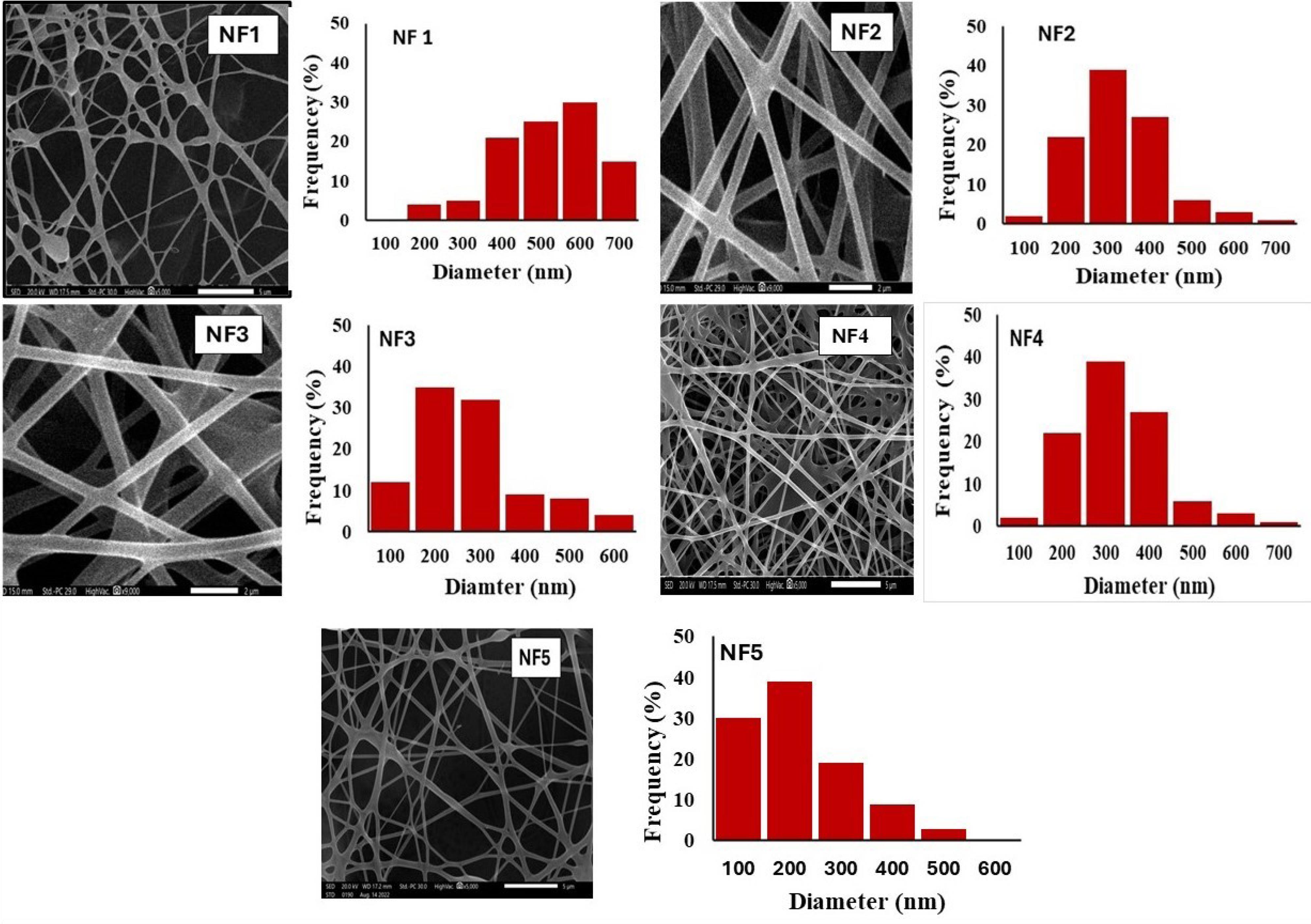
Figure 1.
Scanning electron microscopy images and size distribution histograms of all nanofibers. Images for NF1, NF4 and NF5 have a scale of 5 µm, and that for NF2 and NF3 is 2 µm
.
Scanning electron microscopy images and size distribution histograms of all nanofibers. Images for NF1, NF4 and NF5 have a scale of 5 µm, and that for NF2 and NF3 is 2 µm
Nanofiber NF1 (prepared with no surfactant) showed a mat with interconnected fibers and noticeable beads. This could be due to the relatively low viscosity and high surface tension of the polymeric solution that may have led to the breakup of the liquid jet and formation of beaded fibers.42 The average diameter was computed to be 502.1 ± 90 nm and nanofiber diameter with the highest % frequency was 600 nm.
The average diameters of NF2 through NF5 were significantly (P < 0.05) lower than that of NF1. Addition of surfactants produced more uniform and compact fibers with smooth surfaces and smaller average diameters. For nanofibers prepared using Poloxamer 188, the average diameter of NF2 was 301.8 ± 4 3 nm with the highest % frequency at 300 nm (Figure 1). Further increase in Poloxamer 188 concentration in NF3 (3% w/v) reduced average diameter to 275.5 ± 70 nm, with the highest % frequency at 200 nm (Table 2 and Figure 1).
For mats prepared using Cremophor® RH 40, the average diameter reduced from 290.8 ± 43 nm to 228.3 ± 85 nm when Cremophor® RH 40 concentration increased from 2% (NF4) to 4% w/w (NF5), respectively. The highest % frequency was 300 nm and 200 nm for NF4 and NF5, respectively. It is worth noting that the second highest frequency for NF5 was 100 nm size range. This indicates that the composition of NF5 was optimum for obtaining nanofibers with the smallest fibers diameters and, consequently, the highest length to diameter ratio.
The reduced diameters due to surfactants could be as credited to the slight increase in electrical conductivity and reduction of surface tension as mentioned earlier.23 During electrospinning, the applied high voltage stretches viscoelastic polymeric solution into a cone-like shape known as “Taylor Cone.” When electrical field force overcomes the surface tension, a jet is ejected from this cone’s tip. The presence of surfactant(s) in the polymeric dispersion can induce instability motion of the charged jet and, therefore, produce finer fibers.34 This coincides with previous investigation that reported improved nanofibers quality with reduced diameters by surfactants owing to improved electro-spinnability.43,44
In vitro dissolution studies
Dissolution studies were carried out to investigate the impact of electrospinning on the dissolution of amisulpride. The dissolution profiles of unprocessed amisulpride (control) and different formulations are presented as cumulated amount of the drug released (%) versus time plots (Figure 2). Dissolution parameters were obtained from these profiles and expressed as the percentage of drug released after 5 minutes (Q5), 30 minutes (Q30) and dissolution efficiency (DE%) (Table 2). The dissolution profile of unprocessed amisulpride showed slow and incomplete release. The amount of drug liberated after 5 min (Q5) was 18.2 ± 0.2% with a total dissolution of 60.1 ± 3.7%. A dissolution efficiency of 40.5% reflects poor drug solubility. These unsatisfactory dissolution parameters are mostly due to the crystalline and hydrophobic nature of the drug.1,6
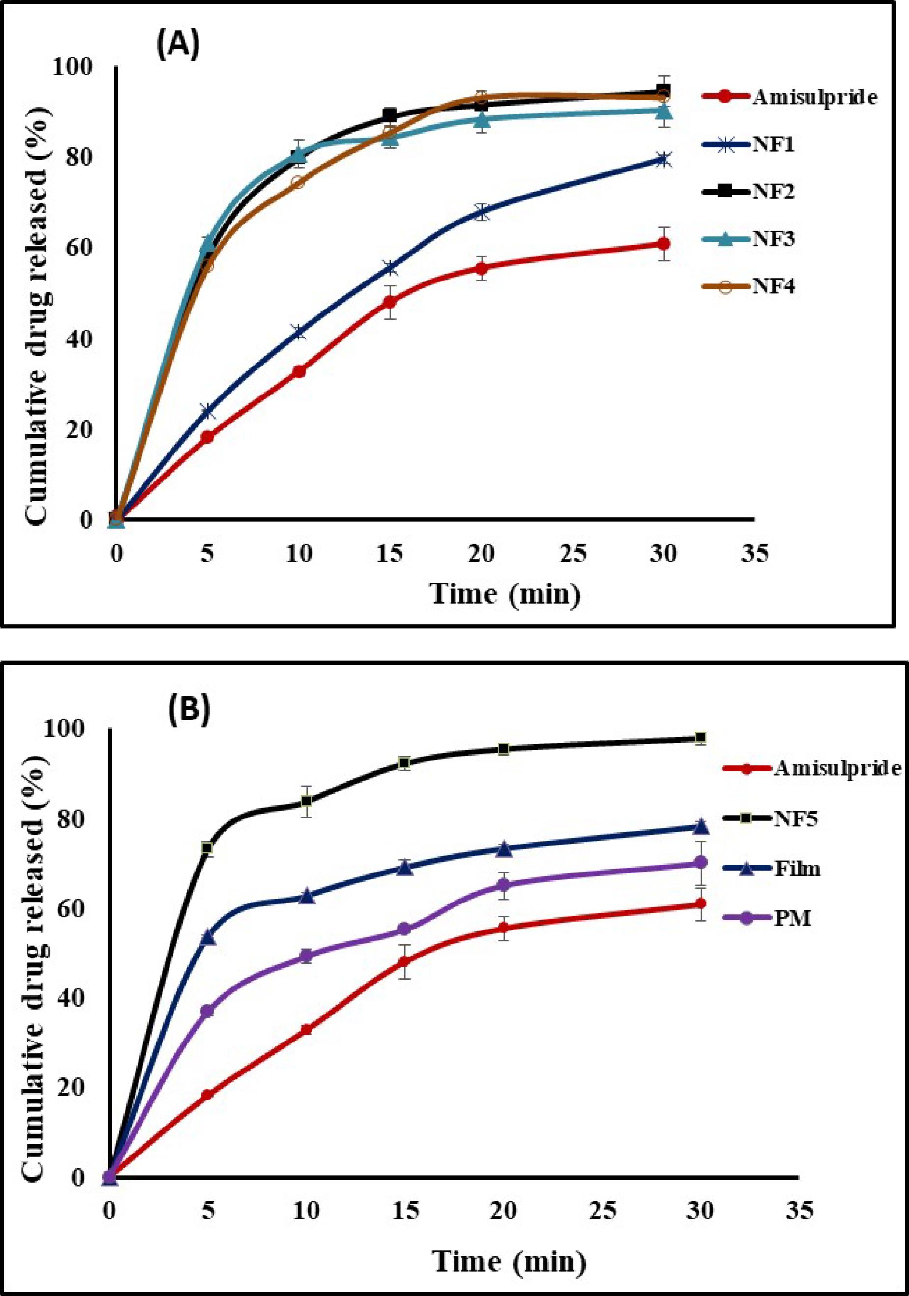
Figure 2.
(A) Dissolution profiles of amisulpride from the unprocessed form and different nanofiber formulations NF1-NF4. (B) Dissolution profiles of amisulpride from the unprocessed form, NF5 mat and the cast Film (n = 3). Detailed formulations are in Table 1
.
(A) Dissolution profiles of amisulpride from the unprocessed form and different nanofiber formulations NF1-NF4. (B) Dissolution profiles of amisulpride from the unprocessed form, NF5 mat and the cast Film (n = 3). Detailed formulations are in Table 1
Regarding NFs, all formulations enhanced (P < 0.05) amisulpride dissolution compared to untreated drug. NF1 mat (prepared without surfactant) showed improvement in Q5 (26.1 ± 2.6%), Q30 (79.5 ± 1.0%) and dissolution efficiency (50.6 ± 1.4%). These results could be accredited to particle size reduction and the significant increase in surface area. Additionally, improved wettability due to the presence of Poly(vinylpyrrolidone) K30 (hydrophilic polymer) could be taken as an additional reason for the observed improvement in drug dissolution.17
As the obtained amisulpride dissolution from NF1 mat was not satisfactory as per our main aim of preparing oro-dispersible film with fast drug release, the inclusion of a surfactant as a ternary component to the polymeric hybrid was investigated. Addition of surfactants considerably improved drug dissolution behavior. Such improvement could be accredited to increased wettability and micellar solubilization posed by the surfactant, in addition to better fibers quality as reflected from SEM images.
Addition of Poloxamer 188 in NF2 mat (at 2% w/v) significantly (P < 0.05) improved drug dissolution compared to unprocessed drug and NF1 mat (Figure 2A). The computed dissolution parameters were 58.2 ± 1.6%, 94.5 ± 3.2% and 87.1 ± 0.9% for Q5, Q30 and dissolution efficiency, respectively. Reduced fibers average diameter with absence of beads increased the surface area, with a subsequent increase in drug dissolution. It is worth noting that, based on the drug loading (Table 2), the concentration of the hydrophilic polymers in NF2 mat is higher than that in NF1. This can further contribute to the superiority of NF2 mat over NF1 mat.
Increasing Poloxamer 188 concentration to 3% w/v in NF3 marginally (P > 0.05) improved dissolution. This was reflected by the similarity value (F2) of 70%, indicating comparable dissolution profiles. Despite the comparable average diameter and drug loading with NF2 mat, it was expected that increasing Poloxamer 188 concentration would improve dissolution from NF3. This unexpected result could be attributed to the reversible thermo-gelation property of Poloxamer 188 that can increase the viscosity of the drug micro-domains.45
With respect to nanofibers containing Cremophor® RH 40, amisulpride dissolution was improved compared to the control and the NF1 mat (Figure 2B). At concentration of 2% w/v, the dissolution parameters of NF4 mat were 55.8 ± 0.8%, 93.0 ± 2.3% and 86.0 ± 0.4% for Q5, Q30 and dissolution efficiency, respectively. These results were comparable to those obtained from NF2 mat as reflected by similarity value of more than 50%. Increasing the concentration of Cremophor® RH 40 (NF5 mat) significantly (P < 0.05) improved drug dissolution over all formulations. This formulation liberated 78.3 ± 0.9% of the loaded amisulpride in the first 5 min with overall release of 98.1 ± 1.3%. The dissolution efficiency was accordingly increased to 90.5 ± 1.0%. A similarity factor (F2) of 47% supports the superiority of NF5 mat over NF4. Such a considerable improvement in drug dissolution could be due to the presence of Cremophor® RH 40, in addition to the vast increment in surface area. The results of nanofibers dissolution indicate that Cremophor® RH 40 (at 4%w/w) was better than Poloxamer 188. This may be explained by taking into consideration the thermos-reversible gelation properties of Poloxamers. At a certain critical temperature (around 24 °C),46 and polymer concentration (about 15% w/v),47 Poloxamer 188 liquid-phase micelles undergo transition to liquid crystalline gel form. Under our experimental conditions, the concentration of Poloxamer 188 in the microenvironment may be enough to form a gel phase that slightly slowed down drug transfer through the diffusion layer.45,48
lower Based on the characterization and in vitro drug dissolution, nanofiber NF5 mat was selected as the optimum formulation.
As film-casting is considered one of the earliest and most adopted methods for film production, film prepared by casting method (Film) was prepared and compared to NF5 mat. The Film was prepared to contain the same components of the optimized nanofibers NF5 mat (Table 1). The Film liberated about 53.7 ± 0.4% of the loaded drug in the first 5 minutes and 78.3 ± 0.9% after 30 min (Figure 2, Table 2). Though these dissolution parameters were significantly higher (P < 0.05) than pure drug, however they were than those for NF5 mat (P > 0.05). The low dissolution efficiency of 74.4%, compared to 90.5% for NF5 mat, together with F2 value of 31% (when comparing the Film to NF5 mat), would indicate the importance of electrospinning technique to achieve our goal of oro-dispersible film with rapid drug release.
Physical state characterization of the optimized nanofibers mat
Differential scanning calorimetry
Thermal behavior of NF5 mat and its individual components were investigated. The obtained thermograms are presented in Figure 3A. The thermogram of unprocessed amisulpride displayed thermal events coincide with the published data.6 The drug exhibited a sharp endothermic peak with an onset of 122.3 °C, endset of 135.8 °C, and the Tm at 127.7 °C. This indicates the crystalline nature of amisulpride. The melting transition was followed by a broad exothermic peak starting at about 240 °C, most probably due to thermal decomposition.
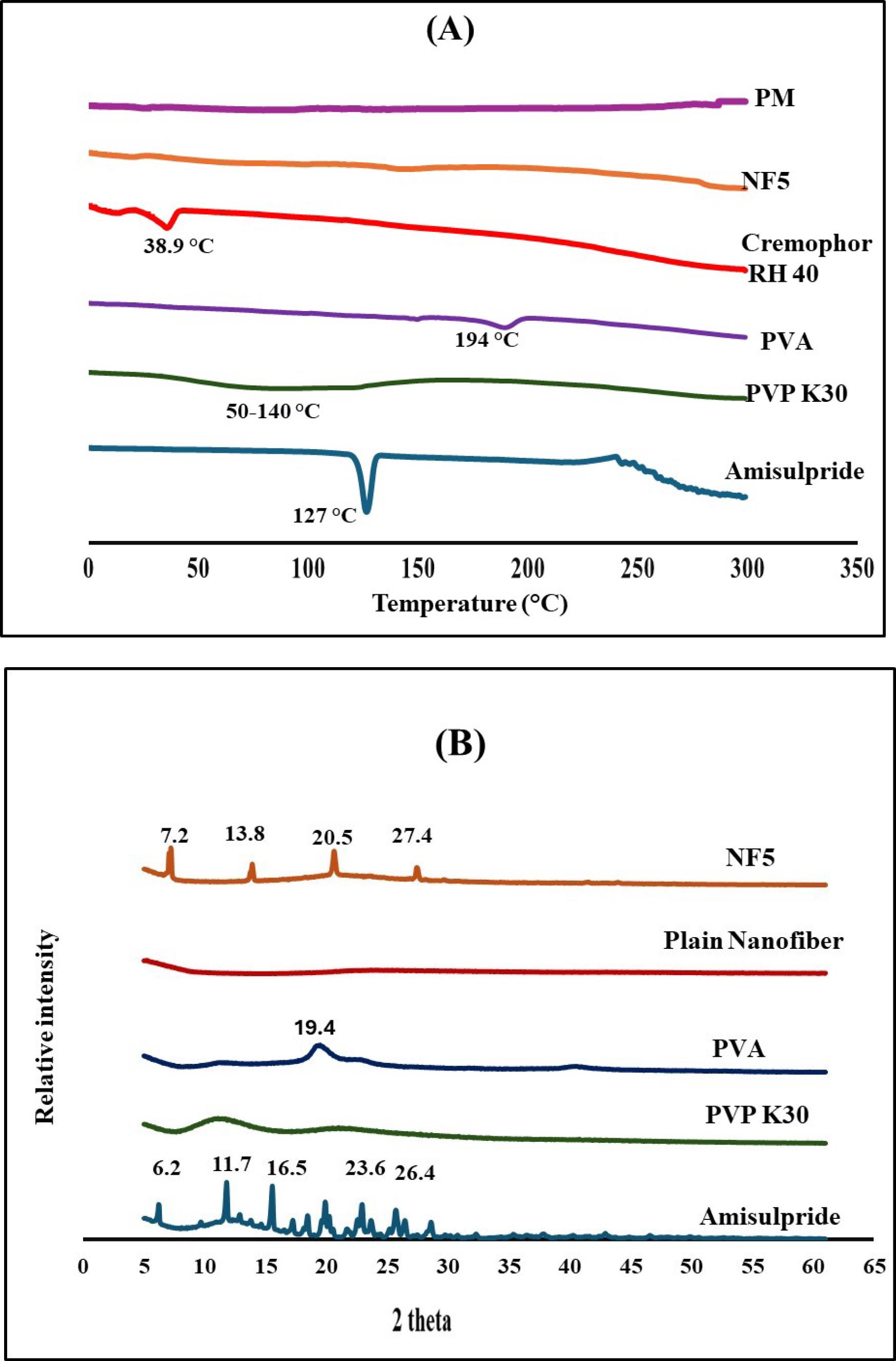
Figure 3.
(A) Differential scanning calorimetry of amisulpride, poly vinyl pyrrolidone(PVP), poly vinyl alcohol (PVA), Cremophor® RH 40 and NF5 mat (B) X-ray diffraction of amisulpride, PVP K30, polyvinyl alcohol (PVA), plain NFs, and NF5 mat
.
(A) Differential scanning calorimetry of amisulpride, poly vinyl pyrrolidone(PVP), poly vinyl alcohol (PVA), Cremophor® RH 40 and NF5 mat (B) X-ray diffraction of amisulpride, PVP K30, polyvinyl alcohol (PVA), plain NFs, and NF5 mat
The thermogram of poly(vinylpyrrolidone) showed a broad endotherm starting from 50 till 140 °C, supposedly due to the loss of adsorbed moisture.49 A broad endothermic peak with onset of 185.6 °C, endset of 203.8 °C and Tm of 194.3 °C was recorded for poly(vinyl alcohol) and represents its melting transition.50 Cremophor® RH 40 thermogram revealed an endothermic peak with Tm of 38.9 °C, indicating its melting transition.51
With respect to NF5 mat, the main melting endotherm of amisulpride disappeared in the thermogram. This could be attributed to either drug solubility in the melted polymer, or reduced drug crystallinity with formation of amorphous form having weak intermolecular forces. Similar findings were reported by other researchers.52,53
Powder X-ray diffraction
PXRD diffraction aimed to further investigate the solid-state properties of the drug before and after processing. The diffractograms of amisulpride, poly(vinylpyrrolidone), poly(vinyl alcohol), NF5 mat as well as plain NF are shown in Figure 3B. The diffractogram of raw amisulpride confirmed its crystalline nature as revealed from the presence of multiple intense diffraction peaks at 2θ values of 6.2, 11.7, 15.5, 19.7, 21.6, 22.7, 23.6, 25.4, 28.4°. Similar PXRD configuration for amisulpride was previously reported.54 For poly(vinylpyrrolidone), the diffractogram revealed diffused pattern with no sharp diffraction peaks indicating its amorphous nature.17,55 PXRD of poly(vinyl alcohol) recorded a broad diffraction peak at 2θ values of 19.48, which is consistent with previously reported data.56
NF5 mat produced product with PXRD pattern that was different from the sum of diffraction patterns of amisulpride and the nanofibers mat forming excipients. Most of the characteristic peaks of the drug and the polymers disappeared. Interestingly, a new sharp diffraction peaks at 2θ values of 7.2, 13.8, 20.5, and 27.4° were recorded. These changes may suggest possible polymorphic transition with different crystalline lattice energy. A new polymorph of amisulpride was previously reported.54 The diffractogram of the plain NF mat showed absence of any diffraction peaks. This would support our supposition that the new peaks were due to polymorphic transition of the drug. However, this supposition requires further future investigations. Surprisingly, this change was not reflected in the DSC, may be due to the very weak crystalline structure of the newly formed polymorph which consumed a small amount of heat that was not detected by the normal DSC. This could explain the improved amisulpride dissolution from NF5 mat compared to cast Film, prepared by solvent casting.
To explain why the suggested drug polymorph showed better dissolution, its crystallinity index was calculated and compared to that of the unprocessed drug. Crystallinity index was found to be 0.031 and 0.012 for unprocessed amisulpride and the new polymorph, respectively. The lower index for the detected polymorph would explain the improved drug dissolution after formulation of nanofibers, in addition to the previously mentioned possible reasons.
Evaluation of the optimized nanofibers mat
The NF5 mat was evaluated regarding surface pH, folding endurance and dissolution time. Surface pH value was measured to elucidate any change in pH value caused by the oro-dispersible film to the buccal mucosa, as shifting in the pH value to more alkaline or acidic value may produce irritation to buccal mucosa. The surface pH of NF5 mat was found to be 7.1 ± 0.5, indicating acceptable value for Oro-mucosal delivery. Buccal films with comparable pH value were similarly developed.31,57 The NF5 mat endured more than 300 folds without cracking, indicating the flexibility of the film. This could be attributed to Cremophor® RH 40 contents that imparted some elasticity to the mat. The film started to disintegrate immediately after the addition of water with complete disintegration after 105 ± 38.0 sec.
In vivo forced swim antidepressant test
Forced swim test is widely accepted as a reliable method to investigate the effect of all major classes of antidepressants. During the test, rodents were forced to swim inside a cylinder containing water, so they are not able to get out and their behavior was scored either swimming and climbing (active behavior) or immobility (passive behavior), the time was reported for both activities.58 A day before conducting the test, a 15-min swimming was performed to highlight the differences in behavior between baseline and the swim test (5-min) after medication treatment on the next day. The reduction in passive behavior and increase in active behavior is usually taken as the antidepressant-like effect.59 Figure 4A shows some of the captured photos for each behavior.
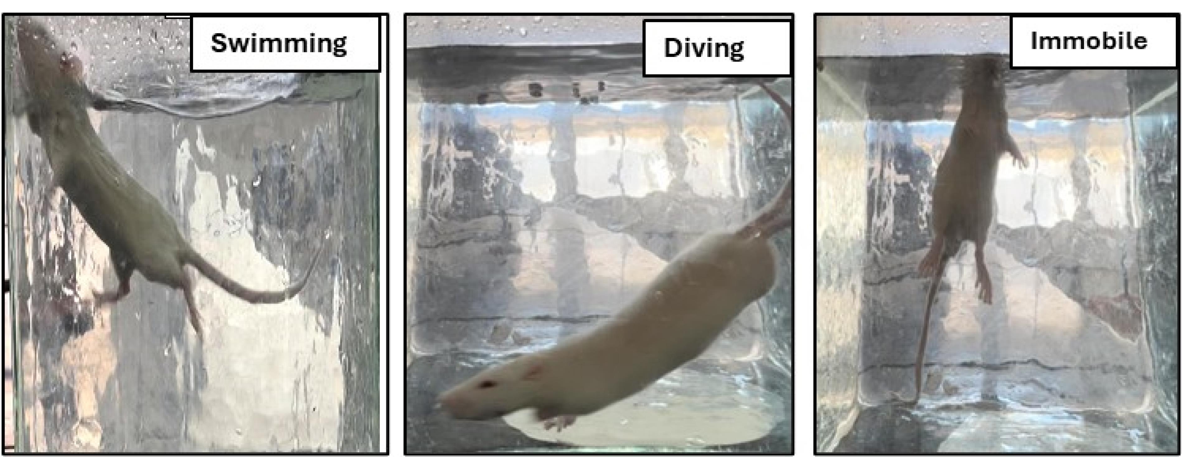
Figure 4.
Photos reflecting active and passive states of rats during the forced swim test
.
Photos reflecting active and passive states of rats during the forced swim test
The effect of the tested formulations on the duration of immobility was calculated and is presented in Table 3, together with the statistical comparison between different groups. The period of immobility is defined as the time during which animals were floating in the water with no struggling and making only the motions required to maintain their heads above water.33
Table 3.
Duration of immobility in rats after administration of distilled water (Placebo n = 6), unprocessed amisulpride suspension (n = 4), and nanofibers formula NF5 mat (n = 4), together with statistical analysis (P value) for comparison of the effect of the administration of NF5 mat in respect to untreated group (placebo) and group received drug suspension
|
|
Treatment
|
Duration of immobility (sec)
|
Percentage reduction in immobility duration
|
P
-value NF5 mat and suspension compared to placebo
|
P
-value NF5 mat compared to suspension
|
| Group 1 |
Placebo |
69.3 ± 5.0 |
- |
- |
- |
| Group 2 |
Drug suspension |
56.8 ± 6.8 |
18.1% |
0.004 |
- |
| Group 3 |
NF5 mat |
44.5 ± 3.2 |
35.8% |
0.000 |
0.004 |
Pretreatment with either unprocessed amisulpride suspension (control group) or NF5 mat (test group) reduced the duration of immobility compared to placebo group (receiving plain water). Statistical comparison (Table 3) implied that administration of NF5 mat and unprocessed drug provided significant (P < 0.05) decrease in the immobility duration compared with the placebo group, with a subsequent increase in the active behavior. There was a reduction in the immobility duration of 18.1% and 35.8% in the positive control and test group, respectively, compared to the placebo group. Compared to drug suspension, NF5 mat reduced immobility time by almost 2-fold. The significant response from NF5 mat coincides with the recorded enhancement in the in vitro drug dissolution study.
Conclusion
-
In this work, amisulpride (BCS Class II) oro-dispersible films were successively prepared using nanofibers technology with the aim of increasing its dissolution.
-
Film matrix composed of poly(vinylpyrrolidone) and poly(vinyl alcohol), with either Poloxamer 188 or Cremophor® RH 40, were successively prepared by electrospinning. Instrumental analysis reflected polymorphic transition of amisulpride after electrospinning.
-
The developed film was in the form of nanofibers which underwent fast liberation of amisulpride. This nanofibrous film was better than traditional film prepared by simple solvent vaporization.
-
The pharmacodynamic properties (i.e. antipsychotic effect) of the optimized nanofiber mat in experimental animals indicated the superiority of nanofiber mat with 2-fold reduction in immobility time relative to unprocessed amisulpride.
-
This research introduces nanofiber technology as a promising tool to prepare oro-dispersible films for buccal delivery of amisulpride with improved antipsychotic effects. Future pharmacokinetics evaluations are recommended.
Competing Interests
None declared.
Ethical Approval
The animal experiments were performed complying with the ethical principles and guidelines for the National Institutes of Health guide for the care and use of laboratory animals. The protocol was reviewed and approved by the research ethics committee, Faculty of Pharmacy, Tanta University, Egypt (Approval code: TP/RE/10/23 p-054).
References
- Scatton B, Claustre Y, Cudennec A, Oblin A, Perrault G, Sanger DJ. Amisulpride: from animal pharmacology to therapeutic action. Int Clin Psychopharmacol 1997; 12 Suppl 2:S29-36. doi: 10.1097/00004850-199705002-00006 [Crossref] [ Google Scholar]
- Gamal W, Fahmy RH, Mohamed MI. Development of novel amisulpride-loaded liquid self-nanoemulsifying drug delivery systems via dual tackling of its solubility and intestinal permeability. Drug Dev Ind Pharm 2017; 43(9):1530-8. doi: 10.1080/03639045.2017.1322607 [Crossref] [ Google Scholar]
- Murali Krishna T, Patel VK, Valluru R, Hiremath JG, Prathapredd D. Solubility enhancement of amisulpride by solid dispersion technique and preparation of fast dissolving tablets. Indo Am J Pharm Res 2012; 1(5):616-28. [ Google Scholar]
- Prajapati Jagruti B, Sawant Krutika K, Dubey B. Oral bioavailability enhancement of amisulpride: complexation and its pharmacokinetics and pharmacodynamics evaluations. Drug Metab Lett 2019; 13(2):132-44. doi: 10.2174/1872312813666191018152226 [Crossref] [ Google Scholar]
- Mankar S, Markad MH, Siddheshwar S, Patel A. Formulation, development, characterization and solubility enhancement of amisulpride. Int J Pharm Sci Nanotechnol 2024; 17(4):7467-74. doi: 10.37285/ijpsn.2024.17.4.4 [Crossref] [ Google Scholar]
- Zhang X, Li J, Rong R, Wang D, Wang D, Yu Y. Enhancing the oral bioavailability of poorly water-soluble amisupiride with solid nanodispersion. J Drug Deliv Sci Technol 2023; 86:104635. doi: 10.1016/j.jddst.2023.104635 [Crossref] [ Google Scholar]
- Sahu MB, Tayde MA. Formulation and evaluation of fast dissolving tablets of amisulpride using co-crystallization technique. Int J Pharm Parm Res 2020; 19(4):181-98. [ Google Scholar]
- Sathali AH, Prakash JC. Formulation and evaluation of amisulpride nanocrystal tablets. Res J Pharm Technol 2015; 8(9):1294-306. doi: 10.5958/0974-360X.2015.00235.8 [Crossref] [ Google Scholar]
- Cetindag E, Pentangelo J, Arrieta Cespedes T, Davé RN. Effect of solvents and cellulosic polymers on quality attributes of films loaded with a poorly water-soluble drug. Carbohydr Polym 2020; 250:117012. doi: 10.1016/j.carbpol.2020.117012 [Crossref] [ Google Scholar]
- Walicová V, Gajdziok J. [Oral films as perspective dosage form]. Ceska Slov Farm 2016;65(1):15-21. [Czech].
- Deepak A, Goyal AK, Rath G. Nanofiber in transmucosal drug delivery. J Drug Deliv Sci Technol 2018; 43:379-87. doi: 10.1016/j.jddst.2017.11.008 [Crossref] [ Google Scholar]
- Subbiah T, Bhat GS, Tock RW, Parameswaran S, Ramkumar SS. Electrospinning of nanofibers. J Appl Polym Sci 2005; 96(2):557-69. doi: 10.1002/app.21481 [Crossref] [ Google Scholar]
- Yu DG, Li JJ, Williams GR, Zhao M. Electrospun amorphous solid dispersions of poorly water-soluble drugs: a review. J Control Release 2018; 292:91-110. doi: 10.1016/j.jconrel.2018.08.016 [Crossref] [ Google Scholar]
- Pelipenko J, Kocbek P, Kristl J. Critical attributes of nanofibers: preparation, drug loading, and tissue regeneration. Int J Pharm 2015; 484(1-2):57-74. doi: 10.1016/j.ijpharm.2015.02.043 [Crossref] [ Google Scholar]
- Huang Y, Song J, Yang C, Long Y, Wu H. Scalable manufacturing and applications of nanofibers. Mater Today 2019; 28:98-113. doi: 10.1016/j.mattod.2019.04.018 [Crossref] [ Google Scholar]
- Kenry Kenry, Lim CT. Nanofiber technology: current status and emerging developments. Prog Polym Sci 2017; 70:1-17. doi: 10.1016/j.progpolymsci.2017.03.002 [Crossref] [ Google Scholar]
- Abdelkader DH, Belal AM, Elkordy EA, Sarhan NI, Essa EA. Fabrication and in-vivo evaluation of polyvinyl pyrrolidone/poloxamer 188 hybrid nanofibers of deflazacort. Int J Pharm 2024; 655:123997. doi: 10.1016/j.ijpharm.2024.123997 [Crossref] [ Google Scholar]
- Illangakoon UE, Gill H, Shearman GC, Parhizkar M, Mahalingam S, Chatterton NP. Fast dissolving paracetamol/caffeine nanofibers prepared by electrospinning. Int J Pharm 2014; 477(1-2):369-79. doi: 10.1016/j.ijpharm.2014.10.036 [Crossref] [ Google Scholar]
- Ullah S, Hashmi M, Hussain N, Ullah A, Sarwar MN, Saito Y. Stabilized nanofibers of polyvinyl alcohol (PVA) crosslinked by unique method for efficient removal of heavy metal ions. J Water Process Eng 2020; 33:101111. doi: 10.1016/j.jwpe.2019.101111 [Crossref] [ Google Scholar]
- Karthikeyan K, Guhathakarta S, Rajaram R, Korrapati PS. Electrospun zein/eudragit nanofibers based dual drug delivery system for the simultaneous delivery of aceclofenac and pantoprazole. Int J Pharm 2012; 438(1-2):117-22. doi: 10.1016/j.ijpharm.2012.07.075 [Crossref] [ Google Scholar]
- Aytac Z, Ipek S, Erol I, Durgun E, Uyar T. Fast-dissolving electrospun gelatin nanofibers encapsulating ciprofloxacin/cyclodextrin inclusion complex. Colloids Surf B Biointerfaces 2019; 178:129-36. doi: 10.1016/j.colsurfb.2019.02.059 [Crossref] [ Google Scholar]
- Zhao J, Koo O, Pan D, Wu Y, Morkhade D, Rana S. The impact of disintegrant type, surfactant, and API properties on the processability and performance of roller compacted formulations of acetaminophen and aspirin. AAPS J 2017; 19(5):1387-95. doi: 10.1208/s12248-017-0104-6 [Crossref] [ Google Scholar]
- Kumar N, Tyagi R. Analysis of the interactions of polyvinylpyrrolidone with conventional anionic and dimeric anionic surfactant. J Dispers Sci Technol 2015; 36(11):1601-6. doi: 10.1080/01932691.2014.981339 [Crossref] [ Google Scholar]
- Bala I, Bhardwaj V, Hariharan S, Kumar MN. Analytical methods for assay of ellagic acid and its solubility studies. J Pharm Biomed Anal 2006; 40(1):206-10. doi: 10.1016/j.jpba.2005.07.006 [Crossref] [ Google Scholar]
- Kaerkitcha N, Chuangchote S, Hachiya K, Sagawa T. Influence of the viscosity ratio of polyacrylonitrile/poly(methyl methacrylate) solutions on core–shell fibers prepared by coaxial electrospinning. Polym J 2017; 49(6):497-502. doi: 10.1038/pj.2017.8 [Crossref] [ Google Scholar]
- Yan T, Shi Y, Zhuang H, Lin Y, Lu D, Cao S. Electrospinning mechanism of nanofiber yarn and its multiscale wrapping yarn. Polymers (Basel) 2021; 13(18):3189. doi: 10.3390/polym13183189 [Crossref] [ Google Scholar]
- Shiekh KA, Liangpanth M, Luesuwan S, Kraisitthisirintr R, Ngiwngam K, Rawdkuen S. Preparation and characterization of bioactive chitosan film loaded with cashew (Anacardium occidentale) leaf extract. Polymers (Basel) 2022; 14(3):540. doi: 10.3390/polym14030540 [Crossref] [ Google Scholar]
- Khan KA. The concept of dissolution efficiency. J Pharm Pharmacol 1975; 27(1):48-9. doi: 10.1111/j.2042-7158.1975.tb09378.x [Crossref] [ Google Scholar]
- Swain RP, Subudhi BB. Effect of Solutol HS 15 and Cremophor RH 40 on dissolution and bioavailability of nateglinide through solid dispersions. Indian J Pharm Educ Res 2022; 56(2):S253-64. [ Google Scholar]
- Maher EM, Ali AM, Salem HF, Abdelrahman AA. In vitro/in vivo evaluation of an optimized fast dissolving oral film containing olanzapine co-amorphous dispersion with selected carboxylic acids. Drug Deliv 2016; 23(8):3088-100. doi: 10.3109/10717544.2016.1153746 [Crossref] [ Google Scholar]
- Elagamy HI, Essa EA, Nouh A, El Maghraby GM. Development and evaluation of rapidly dissolving buccal films of naftopidil: in vitro and in vivo evaluation. Drug Dev Ind Pharm 2019; 45(10):1695-706. doi: 10.1080/03639045.2019.1656734 [Crossref] [ Google Scholar]
- El-Setouhy DA, Abd El-Malak NS. Formulation of a novel tianeptine sodium orodispersible film. AAPS PharmSciTech 2010; 11(3):1018-25. doi: 10.1208/s12249-010-9464-2 [Crossref] [ Google Scholar]
- Rocha BA, Fleischer R, Schaeffer JM, Rohrer SP, Hickey GJ. 17 Beta-estradiol-induced antidepressant-like effect in the forced swim test is absent in estrogen receptor-beta knockout (BERKO) mice. Psychopharmacology (Berl) 2005; 179(3):637-43. doi: 10.1007/s00213-004-2078-1 [Crossref] [ Google Scholar]
- Pawar GR, Agrawal RP, Phadnis P, Paliwal A, Vyas S, Solanki P. Evaluation of antidepressant like property of amisulpride per se and its comparison with fluoxetine and olanzapine using forced swimming test in albino mice. Acta Pol Pharm 2009; 66(3):327-31. [ Google Scholar]
- Diaz DA, Colgan ST, Langer CS, Bandi NT, Likar MD, Van Alstine L. Dissolution similarity requirements: how similar or dissimilar are the global regulatory expectations?. AAPS J 2016; 18(1):15-22. doi: 10.1208/s12248-015-9830-9 [Crossref] [ Google Scholar]
- Shafi H, Reddy DV, Rashid R, Roy T, Kawoosa S, Bader GN. Optimizing the fabrication of electrospun nanofibers of prochlorperazine for enhanced dissolution and permeation properties. Biomater Adv 2024; 158:213773. doi: 10.1016/j.bioadv.2024.213773 [Crossref] [ Google Scholar]
- Mejia ML, Moncada ME, Ossa-Orozco CP. Poly (vinyl alcohol)/silk fibroin/Ag NPs composite nanofibers for bone tissue engineering. Annu Int Conf IEEE Eng Med Biol Soc 2021; 2021:1176-80. doi: 10.1109/embc46164.2021.9629992 [Crossref] [ Google Scholar]
- Dewan M, Adhikari A, Jana R, Chattopadhyay D. Development, evaluation and recent progress of ocular in situ gelling drug delivery vehicle based on poloxamer 407. J Drug Deliv Sci Technol 2023; 88:104885. doi: 10.1016/j.jddst.2023.104885 [Crossref] [ Google Scholar]
- Van Der Hoeven PC, Lyklema J. Electrostatic stabilization in non-aqueous media. Adv Colloid Interface Sci 1992; 42:205-77. doi: 10.1016/0001-8686(92)80024-r [Crossref] [ Google Scholar]
- Dukhin AS, Goetz PJ. How non-ionic “electrically neutral” surfactants enhance electrical conductivity and ion stability in non-polar liquids. J Electroanal Chem (Lausanne) 2006; 588(1):44-50. doi: 10.1016/j.jelechem.2005.12.001 [Crossref] [ Google Scholar]
- Zheng JY, Zhuang MF, Yu ZJ, Zheng GF, Zhao Y, Wang H. The effect of surfactants on the diameter and morphology of electrospun ultrafine nanofiber. J Nanomater 2014; 2014(1):689298. doi: 10.1155/2014/689298 [Crossref] [ Google Scholar]
- Zuo W, Zhu M, Yang W, Yu H, Chen Y, Zhang Y. Experimental study on relationship between jet instability and formation of beaded fibers during electrospinning. Polym Eng Sci 2005; 45(5):704-9. doi: 10.1002/pen.20304 [Crossref] [ Google Scholar]
- Meng H, Wu Q. Surfactant-assisted regulation of polydivinylbenzene nanofibers morphology. Mater Today Chem 2021; 20:100486. doi: 10.1016/j.mtchem.2021.100486 [Crossref] [ Google Scholar]
- Borrego M, Martín-Alfonso JE, Valencia C, Sánchez MC, Franco JM. Influence of surfactants on the electrospinnability of lignin-PVP solutions and subsequent oil structuring properties of nanofiber mats. Polym Bull 2023; 80(6):6885-904. doi: 10.1007/s00289-022-04382-0 [Crossref] [ Google Scholar]
- Balata GF, Zidan AS, Abourehab MA, Essa EA. Rapid disintegrating tablets of simvastatin dispersions in polyoxyethylene-polypropylene block copolymer for maximized disintegration and dissolution. Drug Des Devel Ther 2016; 10:3211-23. doi: 10.2147/dddt.s114724 [Crossref] [ Google Scholar]
- Lin Y, Alexandridis P. Temperature-dependent adsorption of Pluronic F127 block copolymers onto carbon black particles dispersed in aqueous media. J Phys Chem B 2002; 106(42):10834-44. doi: 10.1021/jp014221i [Crossref] [ Google Scholar]
- Sharma PK, Bhatia SR. Effect of anti-inflammatories on Pluronic F127: micellar assembly, gelation and partitioning. Int J Pharm 2004; 278(2):361-77. doi: 10.1016/j.ijpharm.2004.03.029 [Crossref] [ Google Scholar]
- Elkordy AA, Ashoore A, Essa EA. Complexation of naproxen with beta-cyclodextrin with and without poloxamer 407 to enhance drug dissolution. J App Pharm 2012; 3(4):614-27. [ Google Scholar]
- Yadav PS, Kumar V, Singh UP, Bhat HR, Mazumder B. Physicochemical characterization and in vitro dissolution studies of solid dispersions of ketoprofen with PVP K30 and d-mannitol. Saudi Pharm J 2013; 21(1):77-84. doi: 10.1016/j.jsps.2011.12.007 [Crossref] [ Google Scholar]
- Mínguez-García D, Breve N, Capablanca L, Bonet-Aracil M, Díaz-García P, Gisbert-Payá J. Liquid oil trapped inside PVA electrospun microcapsules. Polymers (Basel) 2022; 14(23):5242. doi: 10.3390/polym14235242 [Crossref] [ Google Scholar]
- Mazyed EA, Abdelaziz AE. Fabrication of transgelosomes for enhancing the ocular delivery of acetazolamide: statistical optimization, in vitro characterization, and in vivo study. Pharmaceutics 2020; 12(5):465. doi: 10.3390/pharmaceutics12050465 [Crossref] [ Google Scholar]
- Vasoya JM, Desai HH, Gumaste SG, Tillotson J, Kelemen D, Dalrymple DM. Development of solid dispersion by hot melt extrusion using mixtures of polyoxylglycerides with polymers as carriers for increasing dissolution rate of a poorly soluble drug model. J Pharm Sci 2019; 108(2):888-96. doi: 10.1016/j.xphs.2018.09.019 [Crossref] [ Google Scholar]
- Essa E, Amin M, Sultan A, Arafa M, El Maghraby G, McConville C. Hot melt extrusion for enhanced dissolution and intestinal absorption of hydrochlorothiazide. J Drug Deliv Sci Technol 2023; 88:104895. doi: 10.1016/j.jddst.2023.104895 [Crossref] [ Google Scholar]
- Zhang WP, Chen DY. Crystal structures and physicochemical properties of amisulpride polymorphs. J Pharm Biomed Anal 2017; 140:252-7. doi: 10.1016/j.jpba.2017.03.030 [Crossref] [ Google Scholar]
- Saraf I, Roskar R, Modhave D, Brunsteiner M, Karn A, Neshchadin D. Forced solid-state oxidation studies of nifedipine-PVP amorphous solid dispersion. Mol Pharm 2022; 19(2):568-83. doi: 10.1021/acs.molpharmaceut.1c00678 [Crossref] [ Google Scholar]
- Jeon B, Han D, Yoon G. Piezoelectric characteristics of PVA/DL-alanine polycrystals in d33 mode. iScience 2023; 26(1):105768. doi: 10.1016/j.isci.2022.105768 [Crossref] [ Google Scholar]
- Taelab AA, Elagamy HI, Essa EA. Enhancement of resveratrol dissolution via co-grinding technique: development and evaluation of buccal film. Int J Mod Pharm Res 2022; 6(10):1-17. [ Google Scholar]
- Detke MJ, Rickels M, Lucki I. Active behaviors in the rat forced swimming test differentially produced by serotonergic and noradrenergic antidepressants. Psychopharmacology (Berl) 1995; 121(1):66-72. doi: 10.1007/bf02245592 [Crossref] [ Google Scholar]
- Slattery DA, Cryan JF. Using the rat forced swim test to assess antidepressant-like activity in rodents. Nat Protoc 2012; 7(6):1009-14. doi: 10.1038/nprot.2012.044 [Crossref] [ Google Scholar]