Pharmaceutical Sciences. 31(4):391-402.
doi: 10.34172/PS.025.40953
Research Article
Synthesis, Physicochemical Properties, and Cytotoxicity of Silver Nitrate Nanoparticles of Achillea filipendulina L. Extract in Lung Cancer Cells
Mahtab Jalilzadeh Data curation, Investigation, Visualization, Writing – original draft, 1, # 
Solmaz Asnaashari Conceptualization, Formal analysis, Investigation, Methodology, Project administration, Software, Validation, Writing – original draft, 2, #
Ali Akbar Alizadeh Data curation, Formal analysis, Methodology, Software, Validation, Visualization, Writing – review & editing, 2, *
Siavoush Dastmalchi Project administration, Supervision, 2
Yousef Javadzadeh Conceptualization, Formal analysis, Methodology, Project administration, Supervision, Writing – review & editing, 1, *
Author information:
1Faculty of Pharmacy, Tabriz University of Medical Science, Tabriz, Iran
2Biotechnology Research Center, Tabriz University of Medical Science, Tabriz, Iran
#Equally contributed to this article.
Abstract
Background:
Silver nanoparticles (AgNPs) with varied physicochemical properties have garnered substantial attention in recent years for their applications in imaging, diagnosis, treatment, and drug delivery within nanopharmaceuticals. This study synthesized AgNPs using Achillea filipendulina (A.F) extract as a reducing and stabilizing agent.
Methods:
The synthesized nanoparticles underwent characterization via UV-Vis spectroscopy, FTIR, FESEM, DLS, and zeta potential analysis. The total phenolic content (TPC) method was also employed to estimate the quantity of A. filipendulina extract loaded onto the AgNPs. The antioxidant capacity of A.F-AgNPs was evaluated using the FRAP and DPPH methods and compared to crude extract. The study also investigated the impact of process variables, such as the mixing ratio of reactants, type of solvent, and use of polymers, on the size of A.F-AgNPs to attain optimal sizing. Finally, the cytotoxicity of the nanoparticles was assessed via the MTT assay on the lung adenocarcinoma cell line (A549) and the regular lung fibroblast cell line (MRC-5), comparing it with silver nitrate (AgNO3) and extract.
Results:
The findings indicated a 24% loading of the extract in the synthesized nanoparticles, which mitigated the toxicity of silver nitrate in both cancer and regular cell lines. However, the resulting formulation exhibited more significant toxicity on the cancer cell line. In addition, the in vitro antioxidant assay demonstrated that the extract’s antioxidant potency was four times higher than that of A.F-AgNPs at a concentration of 3.2 mg/mL, which was directly in relationship with drug loading in synthesized nanoparticles.
Conclusion:
The results suggested that polyphenolic compounds or pure phenolic structures act as capping and reducing agents, with more selective effects on cancer cells to minimize side effects on normal lung cells.
Keywords: AgNPs, Nanoparticle, Achillea filipendulina, Antioxidant, Cytotoxicity
Copyright and License Information
© 2025 The Author(s).
This is an open access article and applies the Creative Commons Attribution Non-Commercial License (
http://creativecommons.org/licenses/by-nc/4.0/). Non-commercial uses of the work are permitted, provided the original work is properly cited.
Funding Statement
This research was funded by Faculty of Pharmacy and Biotechnology Research Center, Tabriz University of Medical Sciences (The article is extracted from the student’s dissertation, grant No. 65318).
Introduction
Nanotechnology represents a significant facet of contemporary research within materials science, characterized by the strategic manipulation and resizing of particles. This manipulation results in products with altered chemical, physical, and biological properties, which, due to their enhanced characteristics, have garnered interest across various scientific disciplines, notably in drug delivery applications.1,2 Historically, noble metals and their derivatives have been utilized for their medicinal properties in treating diverse infections. Recent advancements in nanobiotechnology have led to the development of various nanomaterials, including those with extensive applications.3 In particular, silver nanoparticles (AgNPs) stand out in the research landscape due to their unique physicochemical properties and a spectrum of biological activities such as anti-inflammatory, antimicrobial, and anticancer effects.1 These nanoparticles, with a size range of 1-100 nm, exhibit significant alterations in their physical, chemical, and electrical properties, influencing their melting temperature, magnetic behavior, redox potential, and color.4,5
Various methodologies, encompassing both physical and chemical approaches—such as electrochemical techniques,6 microwave irradiation,7 gamma irradiation,8 chemical reduction,9,10 and thermal evaporation11 have been developed to synthesize AgNPs. However, these methods often present industrial and environmental drawbacks, including using toxic chemicals that contribute to environmental pollution and physical techniques that are energy-intensive, costly, and yield low production volumes.12-18 The biosynthesis of nanoparticles emerges as an innovative method to circumvent these limitations, offering a cost-effective, environmentally friendly, and straightforward alternative. This method is characterized by its scalability and simplicity, utilizing capping agents as crucial stabilizers to prevent the formation of oversized nanoparticles and inhibit their aggregation and coagulation during colloidal synthesis.1,2,13-16,18
For the biosynthesis of AgNPs, plant extracts, and microorganisms serve as sources of capping and stabilizing agents.1 Plant extracts, known as green synthesis, are preferred over microorganisms due to their non-toxic, rapid, simple, and cost-effective nature.2,12,14,19 Various plant extracts, mainly medicinal herbs, have recently been explored for the green synthesis of AgNPs, attributed to their biological and therapeutic metabolites.20-22 Within this domain, water is recognized as the ideal solvent for its non-toxic and environmentally safe properties.5
Achillea filipendulina L. (Fernleaf Yarrow), belonging to the Asteraceae family, comprises approximately 130 flowering and perennial species native to Central Asia. This plant has traditionally been employed to treat gout, malaria, arthritis, congestion, and cardiovascular diseases. Investigations into the chemical composition of essential oils and hydro-alcoholic extracts derived from the flowers and leaves of A. filipendulina have revealed a higher concentration of oxygenated monoterpenoids in essential oils. In contrast, hydro-alcoholic extracts are rich in phenolic acids, flavonoids, and bioactive metabolites like tannins and saponins.23,24 Prior research indicates that phenolic acids and flavonoids act as effective capping agents in the synthesis of AgNPs, positioning hydro-alcoholic extracts of this plant as a viable candidate for AgNP synthesis.25
Given the medicinal and therapeutic properties of A. filipendulina, this study pioneers the biological synthesis of AgNP utilizing extracts from the aerial parts of A. filipendulina. Furthermore, the cytotoxic effects of this new formulation were evaluated in the A549 lung cancer cell line.
Methods
Materials
Standard laboratory-grade chemicals/reagents such as silver nitrate (AgNO₃), absolute ethanol, gelatin type A, gallic acid, Folin-Ciocalteu indicator, sodium carbonate powder, and dimethyl sulfoxide (DMSO) were obtained from Merck. Fetal bovine serum (FBS), trypsin, 3-(4,5-dimethylthiazol-2-yl)-2,5-diphenyltetrazolium bromide (MTT), DMEM (Dulbecco’s Modified Eagle Medium), and 2,2-diphenyl-1-picrylhydrazyl (DPPH) were purchased from Sigma Aldrich. Dr. Mojallali provided methanol. All other chemicals used were of analytical grade and obtained from local suppliers.
Plant Extract Preparation
A. filipendulina was gathered from the Tabriz-Marand roadway, East Azerbaijan, Iran, and identified and deposited at the Tabriz Faculty of Pharmacy herbarium (Tbz-fph-136). The extraction method used in this study was maceration. Maceration at room temperature (25 ºC) was carried out three times in actinic glass for 24 hours with 70% ethanol as the solvent. Dried extract powder was obtained by freeze-drying after filtration.26
Biosynthesis of A. filipendulina Silver Nanoparticles (A.F- AgNPs)
Firstly, the solubility of the extract in different solvents, including distilled water, absolute ethanol, and 70% ethanol, was evaluated, and distilled water was selected as the best solvent. In addition, various concentrations of the obtained plant extract were used to synthesize the desired sizes and shapes of A.F-AgNPs. Samples included: (I) 5 mL of the aqueous solution of extract (1.2 mg/mL) was added drop by drop to 22 mL of 1 mM silver nitrate solution, (II) 6 mL of 1.2 mg/mL aqueous solution of extract was added drop by drop to 22 mL of 1 mM silver nitrate solution, (III) 4 mL of 1.56 mg/mL 70% hydro-alcoholic extract solution was added dropwise to 22 mL of 1 mM silver nitrate solution, (IV) 3 mL of absolute ethanol solution of extract (1.96 mg/mL) was added drop by drop to 22 mL of 1 mM silver nitrate solution, (V) 6 mg of gelatin powder was completely dissolved in 10 mL distilled water at 40°C and added to 22 mL of 1 mM silver nitrate aqueous solution, then 6 mL of aqueous extract solution was added drop by drop to them. All the mixtures were continuously stirred (700 rpm) for 15 minutes at room temperature. The mixtures were kept undisturbed until the colorless solution turned brownish-red, revealing the reduction process of Ag+ to Ag0 NPs. The particles were then separated by centrifugation at 12 000 rpm for 20 minutes. Then, the A.F-AgNPs were freeze-dried prior to characterization. The dry powder of AgNPs was stored at -80 °C for the next steps.2,27
UV–Visible Spectroscopy
UV-Vis spectroscopy was employed to confirm the formation of biosynthesized AgNPs. It was utilized to observe simultaneous changes in the color of aqueous dispersion and the UV-Vis spectrum. By gradually adding the extract over time, the color shifted from yellow to brownish-red, and a UV-Vis spectrum was acquired, indicating the biological reduction of silver ions and the formation of AgNPs.14,17 To accomplish this, 1 mL of suspension samples were periodically collected during biosynthesis. Spectroscopy ranged from 300 to 800 nm at different time intervals, such as 5 minutes, 15 minutes, 30 minutes, 1 hour, 2 hours, 4 hours, 8 hours, and 24 hours. The operation was carried out using a UV-Vis spectrophotometer (Agilent, USA).
Fourier Transform Infrared Spectroscopy (FTIR) Analysis
FTIR was conducted to identify functional groups formed during biosynthesis. Samples were prepared by mixing dried A.F-AgNPs with potassium bromide (KBr) in a 1:100 ratio and pressing them into a disc.28 The investigated samples included dried powder of extract, silver nitrate, and A.F-AgNPs. This analysis was performed using the potassium bromide pellet method (FTIR grade).17 and the spectrum was recorded using an infrared spectrometer (Bruker, Germany). IR spectra were collected in the wavelength range from 400-4000 cm⁻¹ and averaged over 9 scans for each measurement with a resolution of 2 cm⁻¹.
Dynamic Light Scattering (DLS) Analysis
DLS is a rapid and straightforward method capable of determining particle size and size distribution based on the Brownian motion of particles. The sample can be a solution or suspension.29 Particle size distribution of NPs was analyzed using Nano-Zeta Sizer (Malvern, UK).
Zeta Potential Measurement
Zeta potential indicates the electrostatic resistance between particles, which determines their surface charge.It is necessary to prepare the sample as a suspension using an ultrasonic device.30 In this study, AgNPs were washed and centrifuged three times, then dispersed in water using ultrasonic dispersion. Analysis was performed using Nano-Zeta Sizer (Malvern, UK).
Scanning Electron Microscopy (SEM) Analysis
SEM is a method used to scan a sample with an electron beam to produce an exaggerated image for determining its surface characteristics and morphology.12,31 This study investigated the surface morphology and exact size of AgNPs using SEM (Tescan, Czech Republic) with an accelerating voltage of 15 kV. An operating pressure of 7 x 10⁻² bar and a current of 20 mA were applied for two minutes during operation.
In-vitro ANTIOXIDANT ASSAYS
The synthesized silver nitrate nanoparticles of A. filipendulina hydro-alcoholic extract were subjected to antioxidant activity evaluation using two different methods: DPPH (2,2-diphenyl-1-picryl hydrazyl) free radical scavenging assay and FRAP (Ferric Reducing Antioxidant Power).32,33
DPPH Free Radical Scavenging Assay
A series of concentrations (7.8–250 µg/mL) of A.F-AgNPs and extract were separately prepared using a serial dilution method. Then, 1 mL of each sample was mixed with 1 mL of DPPH solution (final concentration = 10 µg/mL). The samples were incubated in darkness for 30 minutes. To calculate the reduction of DPPH radical, UV-Vis absorption of all samples was recorded using an Agilent UV-Vis spectrophotometer at 517 nm. The reduction capacity (RC50) was measured according to the following equation:
RC50 = (Absorbance of control-Absorbance of the sample) × 100 / Absorbance of control
FRAP Assay
The method is based on the ability of antioxidants to reduce Fe3+ to Fe2+ in the presence of TPTZ (2,4,6-tripyridyl-s-triazine), forming an intense blue Fe2+–TPTZ complex.34 3 mL of the FRAP solution mixed with 100 µL extract and A.F-AgNPs aqueous solution, both at a concentration of 3.2 mg/mL. The mixtures were incubated in the dark at room temperature for 10 minutes. The absorbance of samples at a wavelength of 593 nm was measured. This operation was carried out by an autoanalyzer (Abbott, USA). The samples’ absorbance was compared to the Ferrous Sulfate standard curve, and the FRAP values were expressed as mmol Fe (II)/mg extract.
Determination of Total Phenolic Content (TPC)
The TPC of the extract and AgNPs was measured by the Folin-Ciocalteu method, and the outcomes were expressed in terms of mg gallic acid equivalent per gram of dry weight of samples (mg GAE g/1 sample dry weight). Aqueous solutions of A.F-AgNPs and extract were prepared at 0.25 mg/mL concentrations. 1 mL of each sample was mixed with 0.2 mL of Folin-Ciocalteu and 1 mL of 2% Na₂CO₃. After 30 minutes of incubation, the absorbance of samples and standards was recorded by a UV-Vis spectrophotometer at 750 nm.34
Cytotoxic Activity
Cytotoxic activity was assessed by the 3-(4,5-dimethylthiazol-2-yl)-2,5-diphenyl-tetrazolium bromide (MTT) assay on A549 lung tumor cell line and MRC-5 lung regular cell line according to the international standards for biological evaluation of medical device (ISO 10993-5:2009). In brief, after cells were counted, they were transferred to 96-well plates at a density of 12,000 cells per well for A549 in columns 6, 7, 8, and 8000 cells per well for MRC-5 in columns 1, 2, and 3. Both cell lines were treated with a series of A.F-AgNPs aqueous suspensions ranging from 6.25 to 400 µL/mL, extract aqueous solutions ranging from 3.125 to 100 µL/mL, and silver nitrate solutions ranging from 4.6875 to 300 µL/mL. A stock concentration (5 mg/mL in phosphate-buffered saline) of MTT solution was prepared, and 20 µL of the solution was added per well and incubated in the dark for 3 hours. After dimethyl sulfoxide (DMSO) was added to dissolve the purple formazan crystals, the plate was shaken in a Heidolph shaker incubator for 20 minutes. The absorbance was recorded by an ELISA reader (Epoch microplate spectrophotometer) at 570 nm. The analysis obtained from the MTT test was performed using GraphPad PRISM (version 2.01) and Excel software.
After calculating the percentage of cell growth using Microsoft Excel software, the percentage of cell growth was calculated with the following formula:
% growth inhibition = (1 - Test V/Max V) × 100
Test V is the number of live cells after loading the sample, and Max V is the number of live cells in the control (negative control). The value of IC50, the sample concentration required for the growth of 50% of the cells, was calculated using the sigmoid dose-response curve by PRISM software and nonlinear regression analysis.35
Results
UV-Vis Spectroscopy
When the reduction of silver ions to A.F-AgNPs occurs, the color starts to change in the solution to brownish-red, indicating the transformation of silver ions into elements. Due to the surface plasmon resonance of A.F-AgNPs, the UV-Vis spectrum shows a peak near 450 nm after 24 hours of incubation, indicating the formation of AgNPs and completion of the reaction (Figure 1).
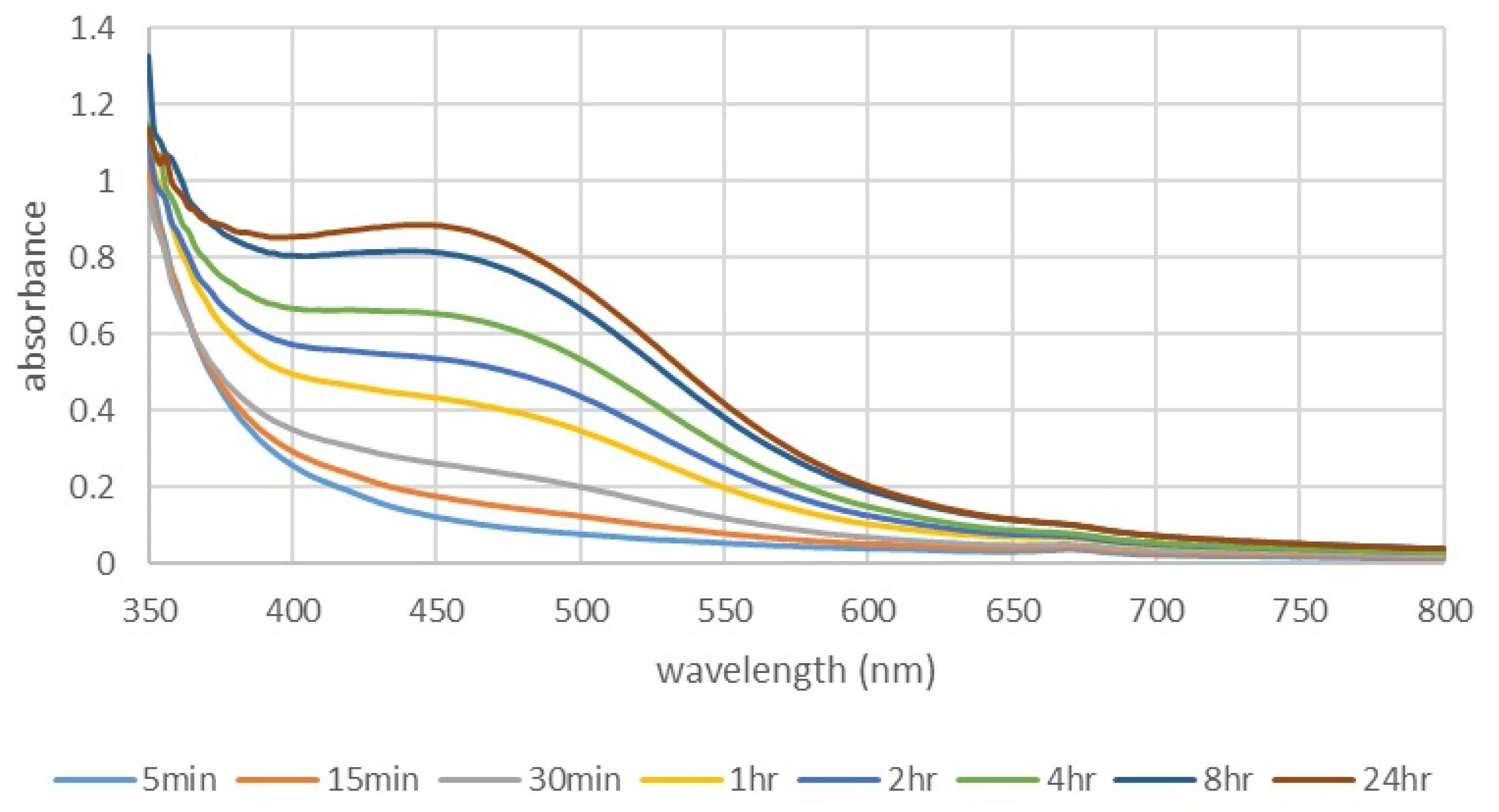
Figure 1.
UV-Vis absorption spectra of A.F-AgNPs at different time intervals. UV-Vis spectrum shows a peak near 450 nm after 24 hours of incubation, indicating the formation of AgNPs.
.
UV-Vis absorption spectra of A.F-AgNPs at different time intervals. UV-Vis spectrum shows a peak near 450 nm after 24 hours of incubation, indicating the formation of AgNPs.
FTIR Analysis
The absorption peaks of the extract spectra displayed at 3413 cm-1, 2926 cm-1, 1620 cm-1 and 1516 cm-1 are attributed to the -OH stretching band of alcohols and phenols, C-H of aliphatic chains or aromatic rings, –C = O stretching band of carbonyl, and C = C stretching band of aromatic rings. The strong band at 3413 cm-1 in A. filipendulina extract was attributed to the -OH stretching band of alcohols and phenols, which shifted to 3420 cm-1 in A.F-AgNPs. The absorption peaks of A.F-AgNPs displayed at 3420 cm-1, 2963–2852 cm-1 and 1621–1638 cm-1 are attributed to the O-H stretching band of alcohol and phenol, C-H of aliphatic chains or aromatic rings, and –C = O stretching band of carbonyl, demonstrating that these bands were formed in biosynthesized NPs (Figure 2).
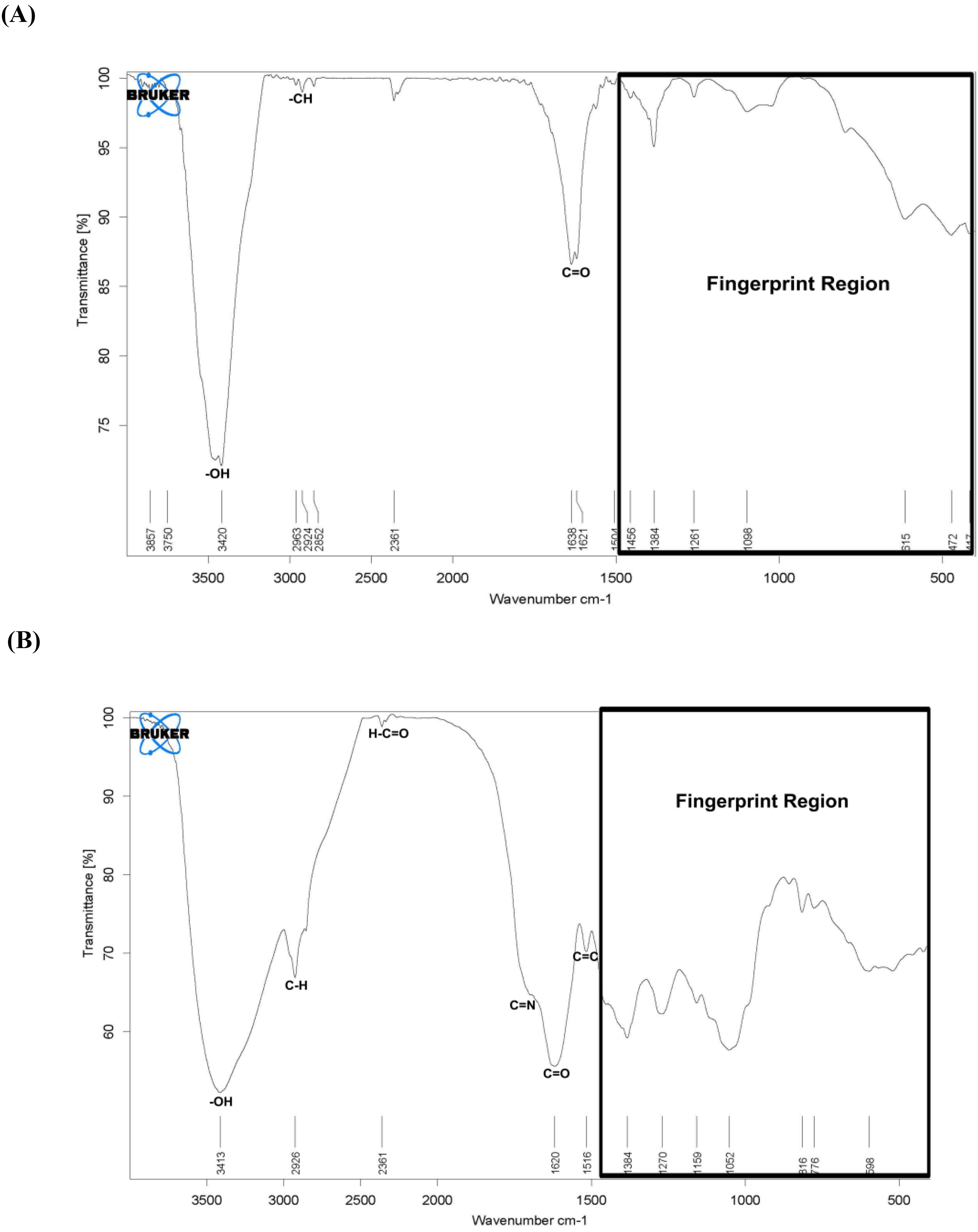
Figure 2.
FTIR spectra of A.F-AgNPs (A) and A. filipendulina extract (B). IR spectra were collected in the wavelength range from 400-4000 cm⁻¹ and averaged over 9 scans for each measurement with a resolution of 2 cm⁻¹.
.
FTIR spectra of A.F-AgNPs (A) and A. filipendulina extract (B). IR spectra were collected in the wavelength range from 400-4000 cm⁻¹ and averaged over 9 scans for each measurement with a resolution of 2 cm⁻¹.
DLS Analysis
NPs A
Based on the number of nanoparticles (NPs), the particle size distribution diagram is a crucial and informative tool for analysis. Only one peak was observed after 24 hours of incubation, indicating a relatively narrow range. The sharper the peak, the closer the particle sizes are to each other. All NPs exhibited a diameter of 48.94 ± 18.93 nm, with an average size of 129.7 nm. The polydispersity index (PDI) was calculated as 0.262, suggesting a narrow size distribution and indicating proximity to the optimal value (Figure 3A). PDI ranges from 0.0 (for a sample exhibiting complete uniformity in size) to 0.1 (for a highly dispersed sample with multiple particle size populations). Values of 0.2 and below are typically deemed optimal for NP materials in size analysis.36,37
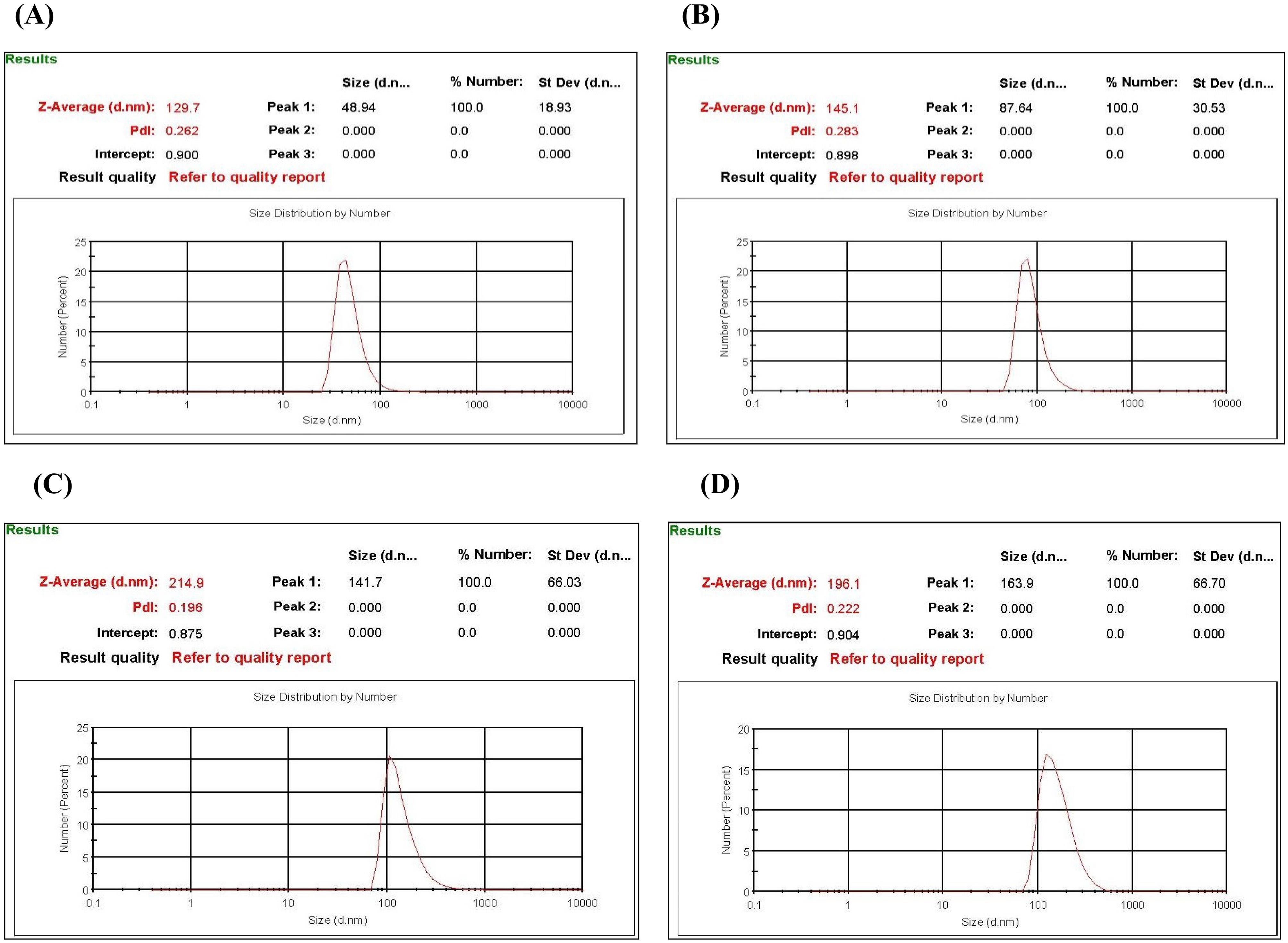
Figure 3.
Particle size analysis of A.F-AgNPs mediated by a 1.6x weight ratio of extract to silver nitrate (A), 2x weight ratio of extract to silver nitrate (B), 70% ethanol extract solution (C), and gelatin (D).
.
Particle size analysis of A.F-AgNPs mediated by a 1.6x weight ratio of extract to silver nitrate (A), 2x weight ratio of extract to silver nitrate (B), 70% ethanol extract solution (C), and gelatin (D).
NPs B
Based on the number of NPs, the particle size distribution diagram exhibited only one peak after 24 hours of incubation, indicating a uniform size distribution. All nanoparticles measured a diameter of 87.64 ± 30.53 nm, with an average size of 145.1 nm. The PDI was 0.283, approaching the optimum value (Figure 3B).
NPs C
Insufficient nanoparticles were synthesized using absolute ethanol as a solvent. After 24 hours of incubation, there was no discernible color change.
NPs D
Based on the number of NPs, the particle size distribution diagram exhibited a single peak after 24 hours of incubation. All nanoparticles measured a diameter of 141.7 ± 66.03 nm, with an average size of 214.9 nm. The PDI was 0.196, representing the optimum value (Figure 3C).
NPs E
Based on the number of NPs, the particle size distribution diagram exhibited a single peak after 24 hours of incubation. All nanoparticles measured a diameter of 163.9 ± 66.70 nm, with an average size of 196.1 nm. The PDI was 0.222, closely approaching the optimum value (Figure 3D).
NPs A After Two Weeks to Estimate the Stability of NPs in Water
Based on the number of NPs, the particle size distribution diagram exhibited a single peak after two weeks of dispersion in water. All nanoparticles measured a diameter of 165.8 ± 61.59 nm, with the average size increasing from 129.7 nm to 200.9 nm. The PDI was 0.245, closely approaching the optimum value.
Zeta Potential of NP A
The zeta potential value determined the surface charge of the NPs. A negative zeta potential of approximately −32.2 mV was recorded in the current study, indicating the high stability of the synthesized NPs (Figure 4). The cell lines used in this study are A549 tumor lung cells and MRC-5 normal lung cells, both non-phagocytic cells. Cellular uptake and cytotoxicity were higher for positively charged surface NPs compared to negatively charged ones in non-phagocytic cells.38-40 Particles with surface charge exhibited more cytotoxicity than neutral nanoparticles.41 The surface charge of A.F-AgNPs is negative, possibly attributed to functionally effective components acting as capping agents in the hydro-alcoholic extract of A. filipendulina.
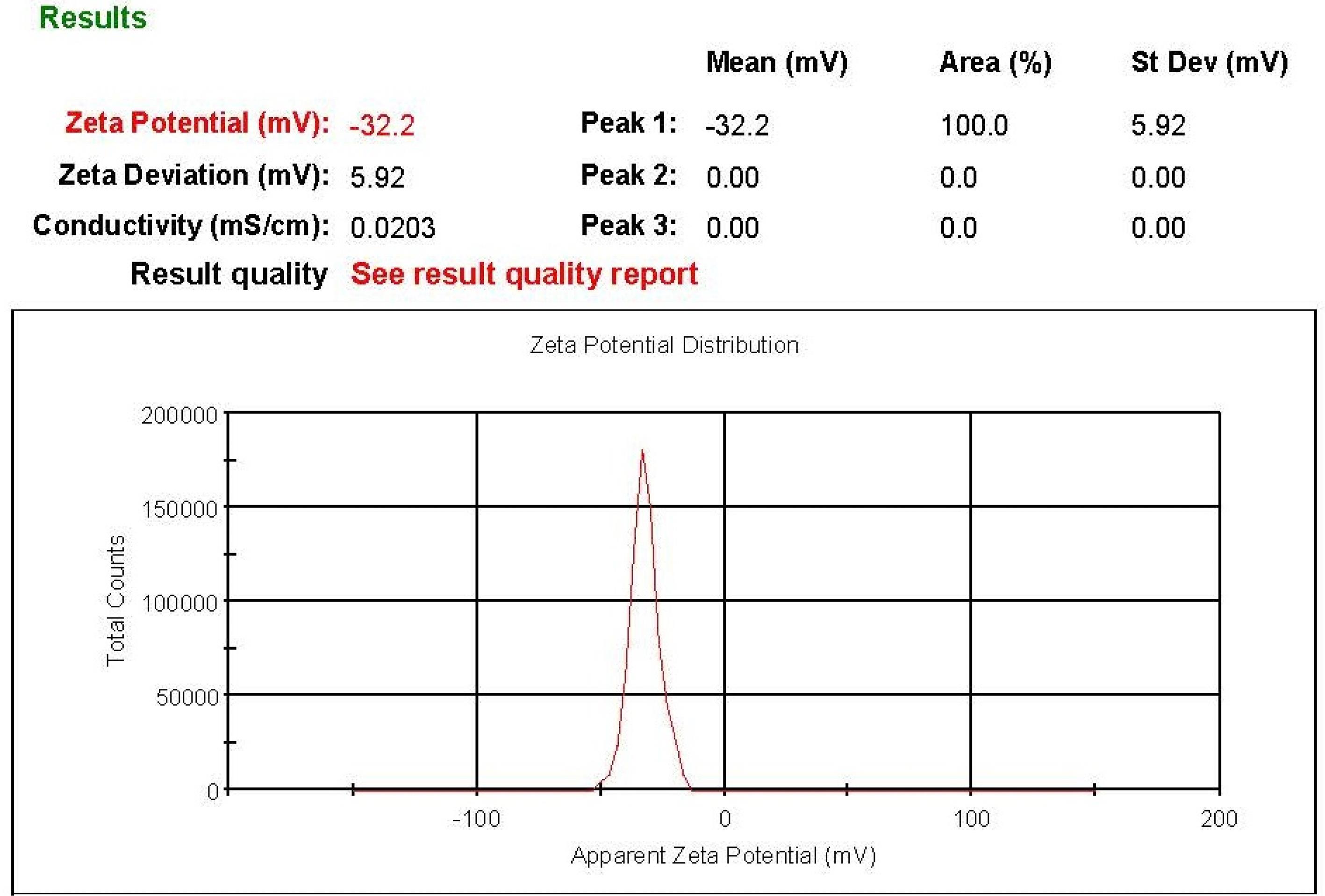
Figure 4.
The Zeta potential of A.F-AgNPs is mediated by a weight ratio of 1.6x.
.
The Zeta potential of A.F-AgNPs is mediated by a weight ratio of 1.6x.
FESEM Analysis
The SEM images revealed that biosynthesized A.F-AgNPs exhibited a spherical morphology with diameters ranging from approximately 38.23 to 55.38 nm, displaying a remarkably narrow diameter distribution (Figure 5).
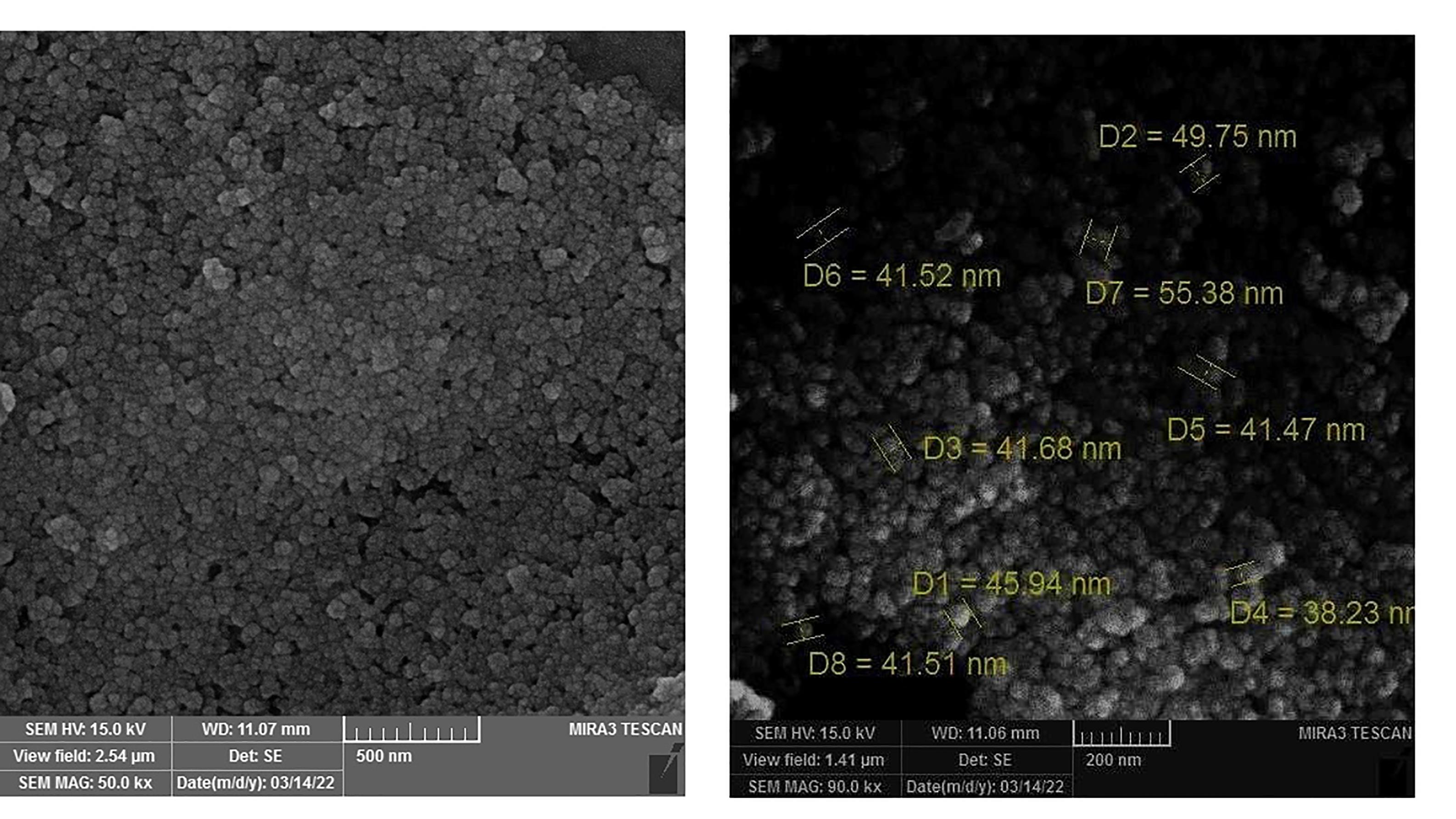
Figure 5.
FE-SEM images of A.F-AgNPs
.
FE-SEM images of A.F-AgNPs
In-vitro Antioxidant Assays
DPPH is a stable free radical compound that readily accepts electrons or hydrogen from AgNPs. The results of free-radical scavenging assays indicate that the reduction percentage increases with the rising concentrations of AgNPs and crude extract.2
The findings demonstrated that the aqueous solution containing A.F-AgNPs and the extract exhibited radical scavenging and antioxidant activities. The RC50 (concentration of antioxidant necessary to scavenge 50% of free radicals) of quercetin, used as the standard compound, was 4 µg/mL, indicating that a lower RC50 value signifies higher antioxidant potency.
Furthermore, the FRAP assay revealed that 3 mL of A.F-AgNPs and extract (3.2 mg/mL) can reduce 1.01 mmol and 4.68 mmol of Fe3+ to Fe2+, respectively. Hence, the extract’s antioxidant potency was four times higher than that of A.F-AgNPs at a concentration of 3.2 mg/mL (Table 1).
Table 1.
Antioxidant results of silver nitrate, AgNPs with extract (A.F-AgNPs), and crude extract
|
Samples
|
DPPH assay
(RC50: µg/mL)
|
FRAP assay (m mol)
|
| Silver nitrate |
N/A |
N/A |
| Silver nanoparticles with extract (A.F-AgNPs) |
181.871 |
1.01 |
| Crude extract |
49.878 |
4.68 |
Total Phenolic Content (TPC)
According to the TPC assay results, the extract’s TPC was determined to be 78.57 mg/g DW (dry weight), while in the AgNPs, it was 18.91 mg/g DW. The FTIR spectra of both the extract and the AgNPs exhibited firm absorption peaks corresponding to the stretching vibration of the hydroxyl (-OH) functional group, indicating the presence of phenolic groups in both. The TPC of the extract was four times that of A.F-AgNPs, corresponding to the proportion of extract loaded onto nanoparticles (24% loading). The results of the TPC assay corroborate those of the antioxidant assay, suggesting that A. filipendulina extract serves as a rich source of phenolic compounds for the biosynthesis of A.F-AgNPs.
Cytotoxic Activity
The cytotoxic activity of A.F-AgNPs, A. filipendulina extract, and silver nitrate was evaluated by assessing the cell viability of MRC-5 normal lung fibroblast cells and A549 lung adenocarcinoma cells following 48 hours of incubation. Figure 6A illustrates that silver nitrate exhibited high cytotoxicity, approaching 100%, even at low concentrations in both cell lines, rendering the calculation of the IC50 (half maximal inhibitory concentration) unfeasible. Conversely, as depicted in Figure 6B, the extract demonstrated no lethality at the tested concentrations, precluding an assessment of its cytotoxic potency. However, the IC50 cannot be computed.
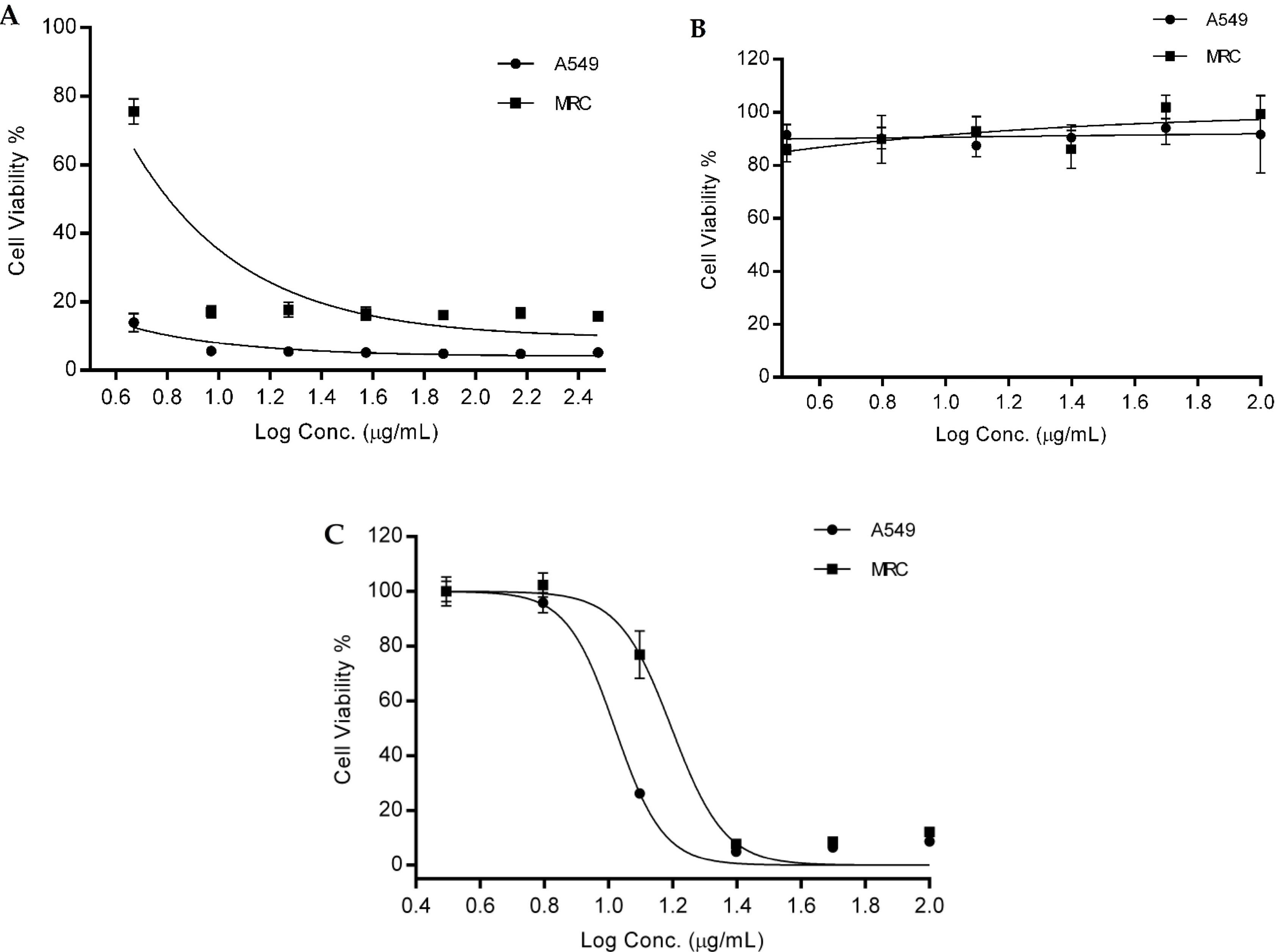
Figure 6.
The percentages of cell viability of MRC-5 normal lung fibroblast cell lines & A549 lung adenocarcinoma cell lines at the presence of various concentrations of silver nitrate (A), A. filipendulina extract (B), and A.F-AgNPs (C)
.
The percentages of cell viability of MRC-5 normal lung fibroblast cell lines & A549 lung adenocarcinoma cell lines at the presence of various concentrations of silver nitrate (A), A. filipendulina extract (B), and A.F-AgNPs (C)
A.F-AgNPs displayed dose-dependent cytotoxicity against both cell lines, as illustrated in Figure 6C The IC50 values of A.F-AgNPs against A549 and the MRC-5 cells were 10.48 ± 1.90 µg/mL and 15.75 ± 1.44 µg/mL, respectively. These results indicated that the cytotoxicity of A.F-AgNPs against A549 tumor cells surpassed that against MRC-5 normal cells, with the cytotoxicity ratio on A549 cells compared to MRC-5 cells estimated at approximately 1.5 times.
Discussion
Today, the prevention and treatment of cancer stand as paramount concerns within the medical sciences. In recent years, extensive research has delved into natural products, notably plants, to discern bioactive compounds and their potential medicinal properties in combating malignancies.12,42 Prior investigations have elucidated the pivotal role of secondary metabolites in plants, augmenting the efficacy of conventional drugs while mitigating their adverse effects.43,44 With advancements in drug delivery systems and the interdisciplinary utilization of biological sciences, including traditional and herbal medicine, alongside cutting-edge biotechnology and nanotechnology tools facilitating the production of green synthesized nanoparticles, a novel strategy has emerged for preventing and treating diseases, including cancer.12
The red-brown color produced by Achillea nanoparticles under the influence of electromagnetic waves occured due to the collective oscillations of the conduction band electrons of A.F-AgNPs because of surface plasmon resonance. According to UV visible spectrum of different concentration of A.F-AgNPs (Figure 1), the appearance of the surface plasmon resonance band with UV-Vis spectrum near 450 nm, absolutely established the successful synthesis of AgNP using the A. filipendulina extract as a reducing agent. This absorbance peak could be used to predict the synthesized A.F-AgNPs.
Previous studies have shown that the silver reduction reaction was attributed to phytochemicals hydroxyl groups.45 FTIR analysis (Figure 2) and TPC assay outcomes confirmed the presence of hydroxyl groups in A. filipendulina extract, which can be responsible for the silver ions reduction reaction in the current report. Moreover, the presence of phenolic structures can be the major classes of plant phytochemicals with antioxidant activities.46 Our studies on the antioxidant properties of the hydroalcoholic extract of A. filipendulina using DPPH and FRAP tests indicated significant antioxidant capacity of extract. In addition, comparing the antioxidant effects of the crude extract and prepared nano-formulation designated the loaded amount of hydroalcoholic extract in prepared nano-formulation (Table 1). A 2020 published study reported significant antioxidant activities of the ethanolic extract of flowers and leaves of A. filipendulina with potent radical scavenging effects equal to 53.93 and 51.70 mg trolox equivalents/grams of sample for DPPH assay and 43.47 and 35.03 mg trolox equivalents/grams of sample for ABTS assay, respectively.24 In addition, a recently published study demonstrated remarkable antioxidant effects of ethanolic extract 18.13 ± 1.53 µg/mL and 432.32 ± 3.43 mg/g using DPPH and FRAP assays, respectively.47
The particle size and surface charge determination of nanoparticles are essential for suitable characterization. DLS and zeta potential measurements have been considered as easy, simple, and reproducible tools for determining particle size and surface charge.48 According to the DLS results in Figure 3, the particle size distribution diagrams showed the prepared A.F-AgNPs with a weight ratio of 1.6 (extract to silver nitrate) water solution was the best nano-formulation for the next cytotoxic studies based on intensity, number, and volume, respectively. The PDI value of the nanoparticles was 0.262 indicating uniformity in the size of the nanoparticles. Although the particle size uniformity (PDI) of prepared nanoparticles with hydroalcoholic solution was slightly better, but the size of them was not optimum. The particle size of A.F-AgNPs prepared with aqueous solution was much smaller (48.94 ± 18.93 nm) and the particle size distribution diagram based on its number is more symmetrical. Moreover, the particle size of A.F-AgNPs prepared in the presence of gelatin was greater and it was not suitable for further studies. As the next step, the prepared A.F-AgNPs with aqueous solution with 1.6 ratio were stored in aqueous solution for two weeks to measure the changes in nanoparticle size and physical stability. The results demonstrated the particle size distribution diagram (based on number) that only one peak at size of 165.8 nm, Z-average of 200.9 nm, and PDI value of 0.245, which showed that after 2 weeks of water exposure of nanoparticles, they have increased in size approximately 2-fold. The average size has increased from 129.7 nm to 200.9 nm. The PDI has decreased from 0.262 to 0.245. Therefore, the uniformity of particle size has increased slightly.
Various nanoparticles with a zeta potential among −10 and + 10 mV are approximately neutral, whereas nanoparticles with zeta potentials of higher than + 30 mV are cationic and the nanoparticles with zeta potentials less than −30 mV are anionic.49
Most cell membranes are negatively charged, therefore zeta potential can affect the tendency of a nanoparticle to penetrate membranes, with cationic particles generally exhibiting greater toxicity in relation to cell wall disruption.49 In addition, one of the general applications of zeta potential data is its relationship to colloidal stability. According to nanoparticle-dispersions guidelines, the zeta potential values of more than + 30 mV and less than -30 mV introduce as highly stable particles in drug delivery literature.49 The result of the evaluation of the zeta potential of prepared A.F-AgNPs (1.6 ratio in aqueous solution) showed the -32.2 ± 5.92 mV (Figures 4 and 5). As mentioned above, the zeta potential value of -32.2 mV confirmed the stability of the nanoparticles. In addition, the uptake of A.F-AgNPs with a negative zeta potential of -32.2 ± 5.92 mV depends on various factors for these particles to be absorbed into lung cells. The negative zeta potential indicates a negative charge on the surface of the particles and they may not be directly absorbed by the cells, but this interaction may be influenced by factors such as particle size. The greater negative zeta potential value may be attributed to the effective functional constituents as capping agents present in the A. filipendulina extract. In this study, the hydro-alcoholic extract of A. filipendulina synthesized AgNPs, investigating their cytotoxic properties on normal and cancerous lung cells. AgNPs have garnered significant attention due to their remarkable physicochemical properties, particularly their potent antimicrobial activity against a spectrum of bacteria, viruses, and fungi.50,51 Moreover, these nanoparticles find application in numerous medical and consumer products.52,53 While it was estimated that approximately 30% of consumer goods contained AgNPs by 2011, these nanoparticles exhibit various toxicities in humans, particularly affecting vital organs such as the lungs.50 Chairuangkitti et al reported that AgNPs induce toxicity in A549 cell lines through ROS (reactive oxygen species)-dependent and ROS-independent pathways. The ROS-dependent pathway, associated with cytotoxicity, involves the generation of ROS within cells, leading to various toxic effects, including disruption of normal MMP and mitochondrial function and induction of cell death, potentially through apoptosis. ROS-independent pathways, manifesting as anti-proliferative activity, operate through cell cycle inhibition with S-phase arrest.50 Despite numerous in vitro and in vivo studies on the therapeutic potential of green synthesized AgNPs for various disorders, many overlook silver nitrate’s toxicity and side effects on lung tissues, commonly used in silver nanoparticle synthesis protocols.54 Fehaid et al. investigated the effects of PVP (polyvinylpyrrolidone)-coated AgNPs and silver nitrate on the inflammatory response following a single intratracheal instillation in rats. Their findings demonstrated that silver ions exhibited more significant toxicity than PVP-coated AgNPs in lung tissue, albeit with higher stability. This discrepancy stemmed from the rapid absorption of silver ions into the bloodstream, leading to acute toxicity dispersed across various tissues, including pulmonary epithelial cells, within 24 hours. In contrast, PVP-coated AgNPs displayed slower absorption and distribution, resulting in subchronic toxicity.54
The cytotoxicity assay results (Figure 6) revealed nearly 100% cytotoxicity for AgNO₃, even at low concentrations, in both A549 cancer cells and MRC-5 normal cells, while the crude plant extract proved safe for both cell lines. Notably, A.F-AgNPs exhibited dose-dependent cytotoxic activity on both cell lines, with an IC50 values of 10.48 ± 1.90 µg/mL for A549 cells and 15.75 ± 1.44 µg/mL for MRC-5 cells. Consequently, A.F-AgNPs demonstrated more significant toxicity toward A549 cancer cells than MRC-5 normal cells. Thus, the A. filipendulina extract utilized in silver nanoparticle synthesis served as a safe capping and reducing agent, attenuating the toxicity of silver nitrate on normal lung cells (MRC-5). It is conceivable that employing plant extracts rich in polyphenolic contents or pure phenolic structures as capping agents may enhance therapeutic effects on cancer cells while minimizing side effects on normal lung cells.
Limitations of Study
The study had several limitations that suggested the necessity of further studies to evaluate the therapeutic potential of A.F-AgNPs more comprehensively:
-
Limited in vitro testing: The cytotoxicity was assessed only on two cell lines (A549, a cancer cell line, and MRC-5, a normal lung fibroblast cell line). Expanding the range of cell lines to include other cancer types and normal tissues could provide a broader understanding of the nanoparticles’ effects.
-
Lack of in vivo evaluation: The study was confined to in vitro methods, which may not fully replicate the complexities of a living organism. In vivo studies in animal models would be necessary to confirm the biological activity, biodistribution, and safety of the synthesized AgNPs.
-
Absence of long-term toxicity and stability data: The study primarily focused on immediate cytotoxicity and antioxidant potential. Long-term studies on the stability of the nanoparticles in biological systems and their possible accumulation or clearance would help assess their viability for therapeutic use.
Conclusion
This study demonstrates the green biosynthesis of AgNPs facilitated by A. filipendulina extract, presenting an uncomplicated, cost-effective, and eco-friendly approach. The metabolites inherent in the extract serve as both capping and reducing agents. The morphology and dimensions of the nanoparticles were characterized using FE-SEM and DLS. AgNPs exhibited significantly lower cytotoxicity in normal and tumor cell lines than silver nitrate. This reduction in cytotoxicity can be attributed to the presence of bioactive compounds from the extract encapsulated within the nanoparticles. FTIR analysis, DPPH scavenging assay, and TPC evaluation indicate that A.F-AgNPs possess antioxidant activity and phenolic functional groups. The higher cytotoxicity observed in tumor cells compared to normal cells is a favorable outcome, suggesting promising potential for further cellular-level research on AgNPs derived from A. filipendulina extract. At concentrations below 10 µg/mL, the cytotoxicity of A.F-AgNPs decreased compared to silver nitrate. However, it is noteworthy that at higher concentrations, the cytotoxicity of A.F-AgNPs approaches that of silver nitrate (100%). Biosynthesized AgNPs hold promise as potential anticancer agents in the biopharmaceutical industry.
Competing Interests
None declared.
Ethical Approval
All experimental procedures were carried out in consistent with the guideline of the Ethics committee of the Tabriz University of Medical Sciences (code: IR.TBZMED.VCR.REC.1400.011).
Acknowledgements
The authors would like to thank Faculty of Pharmacy and Biotechnology Research Center, Tabriz University of Medical Sciences, Tabriz, Iran, for their valuable support (Grant no. 65318). Moreover, we appreciate of the cooperation of Clinical Research Development Unit, Imam Reza General Hospital, Tabriz, Iran in conducting of this research.
References
- Ahmed S, Ahmad M, Swami BL, Ikram S. A review on plants extract mediated synthesis of silver nanoparticles for antimicrobial applications: a green expertise. J Adv Res 2016; 7(1):17-28. doi: 10.1016/j.jare.2015.02.007 [Crossref] [ Google Scholar]
- Salehi S, Shandiz SA, Ghanbar F, Darvish MR, Shafiee Ardestani M, Mirzaie A. Phytosynthesis of silver nanoparticles using Artemisia marschalliana Sprengel aerial part extract and assessment of their antioxidant, anticancer, and antibacterial properties. Int J Nanomedicine 2016; 11:1835-46. doi: 10.2147/ijn.S99882 [Crossref] [ Google Scholar]
- Rai M, Ingle AP, Birla S, Yadav A, Santos CA. Strategic role of selected noble metal nanoparticles in medicine. Crit Rev Microbiol 2016; 42(5):696-719. doi: 10.3109/1040841x.2015.1018131 [Crossref] [ Google Scholar]
- Tomankova K, Horakova J, Harvanova M, Malina L, Soukupova J, Hradilova S. Cytotoxicity, cell uptake and microscopic analysis of titanium dioxide and silver nanoparticles in vitro. Food Chem Toxicol 2015; 82:106-15. doi: 10.1016/j.fct.2015.03.027 [Crossref] [ Google Scholar]
- Yaqoob AA, Umar K, Mohamad Ibrahim MN. Silver nanoparticles: various methods of synthesis, size affecting factors and their potential applications–a review. Appl Nanosci 2020; 10(5):1369-78. doi: 10.1007/s13204-020-01318-w [Crossref] [ Google Scholar]
- Rodríguez-Sánchez L, Blanco MC, López-Quintela MA. Electrochemical synthesis of silver nanoparticles. J Phys Chem B 2000; 104(41):9683-8. doi: 10.1021/jp001761r [Crossref] [ Google Scholar]
- Yin H, Yamamoto T, Wada Y, Yanagida S. Large-scale and size-controlled synthesis of silver nanoparticles under microwave irradiation. Mater Chem Phys 2004; 83(1):66-70. doi: 10.1016/j.matchemphys.2003.09.006 [Crossref] [ Google Scholar]
- Ardjoum N, Shankar S, Chibani N, Salmieri S, Lacroix M. In situ synthesis of silver nanoparticles in pectin matrix using gamma irradiation for the preparation of antibacterial pectin/silver nanoparticles composite films. Food Hydrocoll 2021; 121:107000. doi: 10.1016/j.foodhyd.2021.107000 [Crossref] [ Google Scholar]
- Wang H, Qiao X, Chen J, Ding S. Preparation of silver nanoparticles by chemical reduction method. Colloids Surf A Physicochem Eng Asp 2005; 256(2-3):111-5. doi: 10.1016/j.colsurfa.2004.12.058 [Crossref] [ Google Scholar]
- Gudikandula K, Charya Maringanti S. Synthesis of silver nanoparticles by chemical and biological methods and their antimicrobial properties. J Exp Nanosci 2016; 11(9):714-21. doi: 10.1080/17458080.2016.1139196 [Crossref] [ Google Scholar]
- Kibis LS, Stadnichenko AI, Pajetnov EM, Koscheev SV, Zaykovskii VI, Boronin AI. The investigation of oxidized silver nanoparticles prepared by thermal evaporation and radio-frequency sputtering of metallic silver under oxygen. Appl Surf Sci 2010; 257(2):404-13. doi: 10.1016/j.apsusc.2010.07.002 [Crossref] [ Google Scholar]
- Mousavi B, Tafvizi F, Zaker Bostanabad S. Green synthesis of silver nanoparticles using Artemisia turcomanica leaf extract and the study of anti-cancer effect and apoptosis induction on gastric cancer cell line (AGS). Artif Cells Nanomed Biotechnol 2018; 46(Suppl 1):499-510. doi: 10.1080/21691401.2018.1430697 [Crossref] [ Google Scholar]
- Korbekandi H, Chitsazi MR, Asghari G, Bahri Najafi R, Badii A, Iravani S. Green biosynthesis of silver nanoparticles using Quercus brantii (oak) leaves hydroalcoholic extract. Pharm Biol 2015; 53(6):807-12. doi: 10.3109/13880209.2014.942868 [Crossref] [ Google Scholar]
- Rasheed T, Bilal M, Iqbal HM, Li C. Green biosynthesis of silver nanoparticles using leaves extract of Artemisia vulgaris and their potential biomedical applications. Colloids Surf B Biointerfaces 2017; 158:408-15. doi: 10.1016/j.colsurfb.2017.07.020 [Crossref] [ Google Scholar]
- Khodadadi B, Bordbar M, Nasrollahzadeh M. Achillea millefolium L extract mediated green synthesis of waste peach kernel shell supported silver nanoparticles: application of the nanoparticles for catalytic reduction of a variety of dyes in water. J Colloid Interface Sci 2017; 493:85-93. doi: 10.1016/j.jcis.2017.01.012 [Crossref] [ Google Scholar]
- Bar H, Bhui DK, Sahoo GP, Sarkar P, De SP, Misra A. Green synthesis of silver nanoparticles using latex of Jatropha curcas. Colloids Surf A Physicochem Eng Asp 2009; 339(1-3):134-9. doi: 10.1016/j.colsurfa.2009.02.008 [Crossref] [ Google Scholar]
- Banerjee P, Satapathy M, Mukhopahayay A, Das P. Leaf extract mediated green synthesis of silver nanoparticles from widely available Indian plants: synthesis, characterization, antimicrobial property and toxicity analysis. Bioresour Bioprocess 2014; 1(1):3. doi: 10.1186/s40643-014-0003-y [Crossref] [ Google Scholar]
- Javed R, Zia M, Naz S, Aisida SO, Ain NU, Ao Q. Role of capping agents in the application of nanoparticles in biomedicine and environmental remediation: recent trends and future prospects. J Nanobiotechnology 2020; 18(1):172. doi: 10.1186/s12951-020-00704-4 [Crossref] [ Google Scholar]
- Tripathy A, Raichur AM, Chandrasekaran N, Prathna TC, Mukherjee A. Process variables in biomimetic synthesis of silver nanoparticles by aqueous extract of Azadirachta indica (Neem) leaves. J Nanopart Res 2010; 12(1):237-46. doi: 10.1007/s11051-009-9602-5 [Crossref] [ Google Scholar]
- Kanniah P, Radhamani J, Chelliah P, Muthusamy N, Sathiya Balasingh Thangapandi EJ, Thangapandi JR. Green synthesis of multifaceted silver nanoparticles using the flower extract of Aervalanata and evaluation of its biological and environmental applications. ChemistrySelect 2020; 5(7):2322-31. doi: 10.1002/slct.201903228 [Crossref] [ Google Scholar]
- Narayanan M, Divya S, Natarajan D, Senthil-Nathan S, Kandasamy S, Chinnathambi A. Green synthesis of silver nanoparticles from aqueous extract of Ctenolepisgarcini L and assess their possible biological applications. Process Biochem 2021; 107:91-9. doi: 10.1016/j.procbio.2021.05.008 [Crossref] [ Google Scholar]
- Jebril S, Khanfir Ben Jenana R, Dridi C. Green synthesis of silver nanoparticles using Melia azedarach leaf extract and their antifungal activities: in vitro and in vivo. Mater Chem Phys 2020; 248:122898. doi: 10.1016/j.matchemphys.2020.122898 [Crossref] [ Google Scholar]
- Asghari B, Mafakheri S, Zengin G, Dinparast L, Bahadori MB. In-depth study of phytochemical composition, antioxidant activity, enzyme inhibitory and antiproliferative properties of Achillea filipendulina: a good candidate for designing biologically-active food products. J Food Meas Charact 2020; 14(4):2196-208. doi: 10.1007/s11694-020-00466-5 [Crossref] [ Google Scholar]
- Asnaashari S, Marefat S, Vatankhah AM, Bamdad Moghaddam S, Delazar A, Hamedeyazdan S. Bioactivity assays and phytochemical analysis upon Achillea filipendulina, focusing on xanthine oxidase inhibitory and antimalarial properties. Beni Suef Univ J Basic Appl Sci 2023; 12(1):46. doi: 10.1186/s43088-023-00385-6 [Crossref] [ Google Scholar]
- Pradeep M, Kruszka D, Kachlicki P, Mondal D, Franklin G. Uncovering the phytochemical basis and the mechanism of plant extract-mediated eco-friendly synthesis of silver nanoparticles using ultra-performance liquid chromatography coupled with a photodiode array and high-resolution mass spectrometry. ACS Sustain Chem Eng 2022; 10(1):562-71. doi: 10.1021/acssuschemeng.1c06960 [Crossref] [ Google Scholar]
- Sithisarn P, Supabphol R, Gritsanapan W. Comparison of free radical scavenging activity of Siamese neem tree (Azadirachta indica A Juss var siamensis Valeton) leaf extracts prepared by different methods of extraction. Med Princ Pract 2006; 15(3):219-22. doi: 10.1159/000092185 [Crossref] [ Google Scholar]
- Darroudi M, Bin Ahmad M, Abdullah AH, Ibrahim NA. Green synthesis and characterization of gelatin-based and sugar-reduced silver nanoparticles. Int J Nanomedicine 2011; 6:569-74. doi: 10.2147/ijn.S16867 [Crossref] [ Google Scholar]
- Gavamukulya Y, Maina EN, Meroka AM, Madivoli ES, El-Shemy HA, Wamunyokoli F. Green synthesis and characterization of highly stable silver nanoparticles from ethanolic extracts of fruits of Annona muricata. J Inorg Organomet Polym Mater 2020; 30(4):1231-42. doi: 10.1007/s10904-019-01262-5 [Crossref] [ Google Scholar]
- Murdock RC, Braydich-Stolle L, Schrand AM, Schlager JJ, Hussain SM. Characterization of nanomaterial dispersion in solution prior to in vitro exposure using dynamic light scattering technique. Toxicol Sci 2008; 101(2):239-53. doi: 10.1093/toxsci/kfm240 [Crossref] [ Google Scholar]
- Ezealisiji KM, Noundou XS, Ukwueze SE. Green synthesis and characterization of monodispersed silver nanoparticles using root bark aqueous extract of Annona muricata Linn and their antimicrobial activity. Appl Nanosci 2017; 7(8):905-11. doi: 10.1007/s13204-017-0632-5 [Crossref] [ Google Scholar]
- Goldstein JI, Newbury DE, Michael JR, Ritchie NW, Scott JH, Joy DC. Scanning Electron Microscopy and X-ray Microanalysis. 4th ed. Springer; 2017.
- Asnaashari S, Heshmati Afshar F, Ebrahimi A, Bamdad Moghadam S, Delazar A. Chemical composition and radical scavenging activity of essential oil and methanolic extract of Eremostachysazerbaijanica Rechf from Iran. Res Pharm Sci 2016; 11(2):113-9. [ Google Scholar]
- Salari S, Esmaeilzadeh Bahabadi S, Samzadeh-Kermani A, Yosefzaei F. In-vitro evaluation of antioxidant and antibacterial potential of green synthesized silver nanoparticles using Prosopis farcta fruit extract. Iran J Pharm Res 2019; 18(1):430-55. [ Google Scholar]
- Asgharian P, Delazar A, Vatankhah AM, Javadzadeh M, Asnaashari S. In vitro bioactivity and phytochemical evaluation of extracts from aerial parts of Eremostachys macrophylla Montbr & Auch growing in Iran. Res J Pharmacogn 2017; 4(2):65-73. [ Google Scholar]
- Moulaie S, Mirzaie A, Aliasgari E. Antibacterial and anticancer activities of silver nanoparticles fabricated by the Artemisia scoparia extract against lung cancer cell line (A549). Feyz Med Sci J 2018;22(5):487-96. [Persian].
- Merkus HG. Particle Size Measurements: Fundamentals, Practice, Quality. Springer Science & Business Media; 2009.
- Mudalige T, Qu H, Van Haute D, Ansar SM, Paredes A, Ingle T. Characterization of nanomaterials: tools and challenges. In: Nanomaterials for Food Applications. Elsevier; 2019. p. 313-53. doi: 10.1016/b978-0-12-814130-4.00011-7.
- Bhattacharjee S, de Haan LH, Evers NM, Jiang X, Marcelis AT, Zuilhof H. Role of surface charge and oxidative stress in cytotoxicity of organic monolayer-coated silicon nanoparticles towards macrophage NR8383 cells. Part Fibre Toxicol 2010; 7:25. doi: 10.1186/1743-8977-7-25 [Crossref] [ Google Scholar]
- Goodman CM, McCusker CD, Yilmaz T, Rotello VM. Toxicity of gold nanoparticles functionalized with cationic and anionic side chains. Bioconjug Chem 2004; 15(4):897-900. doi: 10.1021/bc049951i [Crossref] [ Google Scholar]
- Oh WK, Kim S, Choi M, Kim C, Jeong YS, Cho BR. Cellular uptake, cytotoxicity, and innate immune response of silica-titania hollow nanoparticles based on size and surface functionality. ACS Nano 2010; 4(9):5301-13. doi: 10.1021/nn100561e [Crossref] [ Google Scholar]
- Onuma K, Sato Y, Ogawara S, Shirasawa N, Kobayashi M, Yoshitake J. Nano-scaled particles of titanium dioxide convert benign mouse fibrosarcoma cells into aggressive tumor cells. Am J Pathol 2009; 175(5):2171-83. doi: 10.2353/ajpath.2009.080900 [Crossref] [ Google Scholar]
- Amjad E, Sokouti B, Asnaashari S. A systematic review of anti-cancer roles and mechanisms of kaempferol as a natural compound. Cancer Cell Int 2022; 22(1):260. doi: 10.1186/s12935-022-02673-0 [Crossref] [ Google Scholar]
- Seca AM, Pinto D. Plant secondary metabolites as anticancer agents: successes in clinical trials and therapeutic application. Int J Mol Sci 2018; 19(1):263. doi: 10.3390/ijms19010263 [Crossref] [ Google Scholar]
- Asnaashari S, Amjad E, Sokouti B. Synergistic effects of flavonoids and paclitaxel in cancer treatment: a systematic review. Cancer Cell Int 2023; 23(1):211. doi: 10.1186/s12935-023-03052-z [Crossref] [ Google Scholar]
- Ahn EY, Jin H, Park Y. Green synthesis and biological activities of silver nanoparticles prepared by Carpesiumcernuum extract. Arch Pharm Res 2019; 42(10):926-34. doi: 10.1007/s12272-019-01152-x [Crossref] [ Google Scholar]
- Lakshmi CH, Raju BD, Madhavi T, Sushma NJ. Identification of bioactive compounds by FTIR analysis and in vitro antioxidant activity of Clitoriaternatea leaf and flower extracts. Indo Am J Pharm Res 2014; 4(9):3894-903. [ Google Scholar]
- Tunç T, Akın Ş, Aykaç O, Hepokur C, Duran S, Özpınar H. Antioxidant, antimicrobial, and anticancer effects of Achillea filipendulina L against colon cancer. Asian Pac J Trop Biomed 2024; 14(12):540-50. doi: 10.4103/apjtb.apjtb_515_24 [Crossref] [ Google Scholar]
- Bhattacharjee S. DLS and zeta potential - what they are and what they are not?. J Control Release 2016; 235:337-51. doi: 10.1016/j.jconrel.2016.06.017 [Crossref] [ Google Scholar]
- Clogston JD, Patri AK. Zeta potential measurement. In: McNeil SE, ed. Characterization of Nanoparticles Intended for Drug Delivery. Totowa, NJ: Humana Press; 2011. p. 63-70. doi: 10.1007/978-1-60327-198-1_6.
- Chairuangkitti P, Lawanprasert S, Roytrakul S, Aueviriyavit S, Phummiratch D, Kulthong K. Silver nanoparticles induce toxicity in A549 cells via ROS-dependent and ROS-independent pathways. Toxicol In Vitro 2013; 27(1):330-8. doi: 10.1016/j.tiv.2012.08.021 [Crossref] [ Google Scholar]
- Szmyd R, Goralczyk AG, Skalniak L, Cierniak A, Lipert B, Filon FL. Effect of silver nanoparticles on human primary keratinocytes. Biol Chem 2013; 394(1):113-23. doi: 10.1515/hsz-2012-0202 [Crossref] [ Google Scholar]
- Shanmuganathan R, Karuppusamy I, Saravanan M, Muthukumar H, Ponnuchamy K, Ramkumar VS. Synthesis of silver nanoparticles and their biomedical applications - a comprehensive review. Curr Pharm Des 2019; 25(24):2650-60. doi: 10.2174/1381612825666190708185506 [Crossref] [ Google Scholar]
- Yoo M, Kim HK, Kim S, Tentzeris M, Lim S. Silver nanoparticle-based inkjet-printed metamaterial absorber on flexible paper. IEEE Antennas Wirel Propag Lett 2015; 14:1718-21. doi: 10.1109/lawp.2015.2420712 [Crossref] [ Google Scholar]
- Fehaid A, Hamed MF, Abouelmagd MM, Taniguchi A. Time-dependent toxic effect and distribution of silver nanoparticles compared to silver nitrate after intratracheal instillation in rats. Am J Nanomater 2016; 4(1):12-9. doi: 10.12691/ajn-4-1-3 [Crossref] [ Google Scholar]